- Ophthalmic artery
-
Artery: Ophthalmic artery 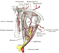
The ophthalmic artery and its branches. 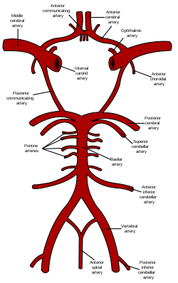
Circle of Willis (Ophthalmic artery labeled at upper right) Latin arteria ophthalmica Gray's subject #146 568 Source Internal carotid Branches Lacrimal artery
Supraorbital artery
Posterior ethmoidal artery
Anterior ethmoidal artery
Internal palpebral artery
Supratrochlear artery
Dorsal nasal artery
Posterior ciliary arteries
Long posterior ciliary arteries
Short posterior ciliary arteries
Anterior ciliary artery
Central retinal artery
Superior muscular artery
Inferior muscular arteryVein superior ophthalmic, inferior ophthalmic MeSH Ophthalmic+Artery The ophthalmic artery (OA) is the first branch of the internal carotid artery distal to the cavernous sinus. Branches of the OA supply all the structures in the orbit as well as some structures in the nose, face and meninges. Occlusion of the OA or its branches can produce sight-threatening conditions.
Contents
Course and branches
Course from internal carotid to orbit
The OA emerges from the internal carotid artery usually just after the latter emerges from the cavernous sinus although in some cases, the OA branches just before the internal carotid exits the cavernous sinus. The OA arises from the internal carotid along the medial side of the anterior clinoid process and runs anteriorly passing through the optic canal with and inferolaterally to the optic nerve. Here, it should be noted that the ophthalmic artery can also pass superiorly to the optic nerve in a minority of cases [1]. In the posterior third of the cone of the orbit, the ophthalmic artery turns sharply medially to run along the medial wall of the orbit.
Central retinal artery
The central retinal artery is the first, and one of the smaller branches of the OA and runs in the dura mater inferior to the optic nerve. About 12.5mm (0.5 inches) posterior to the globe, the central retinal artery turns superiorly and penetrates the optic nerve continuing along the center of the optic nerve entering the eye to supply the inner retinal layers.
Lacrimal artery
The next branch of the OA is the lacrimal artery, one of the largest, arises just as the OA enters the orbit and runs along the superior edge of the lateral rectus muscle to supply the lacrimal gland, eyelids and conjunctiva.
Posterior ciliary arteries
The OA then turns medially giving off 1 to 5 posterior ciliary arteries (PCA) that subsequently branch into the long and short posterior ciliary arteries (LPCA and SPCA respectively) which perforate the sclera posteriorly in the vicinity of the optic nerve and macula to supply the posterior uveal tract. In the past, anatomists made little distinction between the posterior ciliary arteries and the short and long posterior ciliary arteries often using the terms synonymously. However, recent work by Hayreh has shown that there is both an anatomic and clinically useful distinction[2]. The PCAs arise directly from the OA and are end arteries which is to say no PCA or any of its branches anastomose with any other artery. Consequently, sudden occlusion of any PCA will produce an infarct in the region of the choroid supplied by that particular PCA. Occlusion of a short or long PCA will produce a smaller choroidal infarct within the larger area supplied by the specific parent PCA.
Muscular branches
The OA continues medially the superior and inferior muscular branches arise either from the OA or a single trunk from the OA subsequently divides into superior and inferior branches to supply the extraocular muscles.
Supraorbital artery
The supraorbital artery branches from the OA as it passes over the optic nerve. The supraorbital artery passes anteriorly along the medial border of the superior rectus and levator palpebrae and through the supraorbital foramen to supply muscles and skin of the forehead.
Ethmoidal arteries
After reaching the medial wall of the orbit, the OA again turns anteriorly. The posterior ethmoidal artery enters the nose via the posterior ethmoidal canal and supplies the posterior ethmoidal sinuses and enters the skull to supply the meninges.
The OA continues anteriorly, giving off the anterior ethmoidal artery which enters the nose after traversing the anterior ethmoidal canal and supplies the anterior and middle ethmoidal sinuses as well as the frontal sinus and also enters the cranium to supply the meninges.
Medial palpebral arteries
The OA continues anteriorly to the trochlea where the medial palpebral arteries (superior and inferior) arise and supply the eyelids.
Terminal branches
The OA terminates in two branches, the supratrochlear (or frontal) artery and the dorsal nasal artery. Both exit the orbit medially to supply the forehead and nose.
Classification of ophthalmic artery branches
Because of the obvious importance of the ocular globe branches of the ophthalmic artery are often subdivided into two groups: those that supply the eyeball (ocular group) and those that supply nonocular orbital structures (orbital group). http://education.yahoo.com/reference/gray/subjects/subject/146
Orbital group
The orbital group, distributing vessels to the orbit and surrounding parts, includes:
- Lacrimal artery
- Supraorbital artery
- Posterior ethmoidal artery
- Anterior ethmoidal artery
- Internal palpebral artery
- Frontal artery, also called the Supratrochlear artery
- Dorsal nasal artery
Ocular group
The ocular group, distributing vessels to the eye and its muscles, includes:
- Posterior ciliary arteries
- Long posterior ciliary arteries
- Short posterior ciliary arteries
- Anterior ciliary artery
- Central retinal artery
- Superior muscular artery
- Inferior muscular artery
Structures supplied by ophthalmic artery
Branches of the ophthalmic artery supply:
- Frontal belly of the occipitofrontalis muscle
- Inferior oblique muscle
- Inferior rectus muscle
- Lacrimal gland
- Lateral rectus muscle
- Levator palpebrae superioris muscle
- Medial rectus muscle
- Nasalis muscle
- Procerus muscle
- Superior oblique muscle
- Superior rectus muscle
Occlusion
Further information: Ocular ischemic syndromeSevere occlusion of the ophthalmic artery causes ocular ischemic syndrome. As with central retinal artery occlusions, ophthalmic artery occlusions may result from systemic cardiovascular diseases; however, a cherry-red spot is typically absent and the vision is usually worse. Amaurosis fugax is a temporary loss of vision that occurs in two conditions which cause a temporary reduction in ophthalmic artery pressure: orthostatic hypotension and positive acceleration.[3]
Even complete occlusion of the ophthalmic artery may possibly leave the eye without symptoms, probably because of circulatory anastomoses [4]
Additional images
-
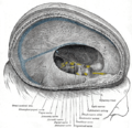
Dura mater and its processes exposed by removing part of the right half of the skull, and the brain.
References
- ^ http://www.medscape.com/viewarticle/562682_4
- ^ Hayreh, SS. "Posterior Ciliary Artery Circulation In Health and Disease" Investigative Ophthalmology and Visual Science 2004 Mar;45(3):749-757. PMID 14985286
- ^ Phelps GK, Phelps CD. "Blood pressure and pressure amaurosis." Investigative Ophthalmology and Visual Science 1975 Mar;14(3):237-40. PMID 1116922
- ^ A case of ophthalmic artery occlusion without manifestation of ocular ischemic syndrome. Authors;SHIMABUKURO MIKIKO(Izumisano City Hosp.) OJI MASATO(Osaka Univ., Med. Sch.) AOMATSU ICHIKO(Osaka Police Hosp.) FUKUI TAKEHIRO(Osaka Police Hosp.) TSUKAMOTO HIROKO(Osaka Police Hosp.) TANAKA YASUO(Osaka Police Hosp.) NISHIKAWA NORIKIYO(Osaka Police Hosp.) KITANISHI KUNIKO(Shiritsuizumiotsubyoin) OZAKI TOSHIYA(Kojunkaiobpkurinikku). Journal Title;Japanese Journal of Clinical Ophthalmology. Journal Code:Z0515B. ISSN:0370-5579. VOL.54;NO.1;PAGE.97-101(2000)
See also
External links
List of arteries of head and neck (TA A12.2.05–08, GA 6.549) CC cervical branches (ascending palatine, tonsillar, submental, glandular) · facial branches (inferior labial, superior labial / nasal septum, lateral nasal, angular)1st part / mandibular2nd part / pterygoid3rd part / pterygopalatinecavernous/
ophthalmicorbital group:anterior ethmoidal (anterior septal, anterior lateral nasal, anterior meningeal) · posterior ethmoidal · lacrimal (lateral palpebral) · medial palpebral · terminal (supraorbital, supratrochlear, dorsal nasal)
ocular group: central retinal · ciliary (short posterior, long posterior, anterior) · hypophysial (superior, inferior)ACA (anterior communicating, medial striate) · MCA (anterolateral central, Orbitofrontal artery, Prefrontal artery, Superior terminal branch, Inferior terminal branch, Anterior temporal branch) · posterior communicating · anterior choroidalSC Categories:- Arteries of the head and neck
- Cardiovascular system stubs
- Eye stubs


Wikimedia Foundation. 2010.