- Dockerin
-
Dockerin domain 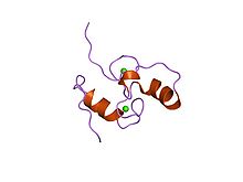
Structure of the Dockerin type I domain from C. thermocellum cellulosome. Identifiers Symbol Dockerin_1 Pfam PF00404 InterPro IPR018242 PROSITE PDOC00416 SCOP 1daq Available protein structures: Pfam structures PDB RCSB PDB; PDBe PDBsum structure summary Dockerin is a protein domain found in the Cellulosome cellular structure. It is part of endoglucanase enzymes. The dockerin's binding partner is the cohesin domain. This interaction is essential to the construction of the Cellulosome complex (also known as a Scaffoldin). The Dockerin domain has two in-tandem repeats of a non-EF hand calcium binding motif. Each motif is characterized by a loop-helix structure.[1] The three dimensional structure of dockerin has been determined in solution,[2] as well as in complex with Cohesin.[3]
There are three types of Dockerin domains: I, II and III which bind to Cohesin Type I, Cohesin Type II and Cohesin Type III respectively. A type I dockerin domain is 65-70 residues long.[4] The binding specificity of Type I interaction was well studied by structural and mutagenesis studies. Type II interaction is less well characterized.[5]
See also
References
- ^ SCOP 63447
- ^ PDB 1DAQ; Lytle BL, Volkman BF, Westler WM, Heckman MP, Wu JH (March 2001). "Solution structure of a type I dockerin domain, a novel prokaryotic, extracellular calcium-binding domain". J. Mol. Biol. 307 (3): 745–53. doi:10.1006/jmbi.2001.4522. PMID 11273698.
- ^ PDB 1OHZ; Carvalho AL, Dias FM, Prates JA, Nagy T, Gilbert HJ, Davies GJ, Ferreira LM, Romão MJ, Fontes CM (November 2003). "Cellulosome assembly revealed by the crystal structure of the cohesin–dockerin complex". Proc. Natl. Acad. Sci. U.S.A. 100 (24): 13809–14. doi:10.1073/pnas.1936124100. PMC 283503. PMID 14623971. http://www.pubmedcentral.nih.gov/articlerender.fcgi?tool=pmcentrez&artid=283503.
- ^ InterPro: IPR016134
- ^ Adams JJ, Webb BA, Spencer HL, Smith SP (February 2005). "Structural characterization of type II dockerin module from the cellulosome of Clostridium thermocellum: calcium-induced effects on conformation and target recognition". Biochemistry 44 (6): 2173–82. doi:10.1021/bi048039u. PMID 15697243.
External links
Protein Structure:
- Lytle BL, Volkman BF, Westler WM, Heckman MP, Wu JH (2001). "Solution structure of a type I dockerin domain, a novel prokaryotic, extracellular calcium-binding domain". J Mol Biol 307 (3): 745–753. doi:10.1006/jmbi.2001.4522. PMID 11273698.
- Bayer EA, Shimon LJ, Shoham Y, Lamed R (1998). "Cellulosomes-structure and ultrastructure". J Struct Biol 124 (2–3): 221–234. doi:10.1006/jsbi.1998.4065. PMID 10049808.
Specificity Characterization:
- Haimovitz R, Barak Y, Morag E, Voronov-Goldman M, Shoham Y, Lamed R, Bayer EA (2008). "Cohesin-dockerin microarray: Diverse specificities between two complementary families of interacting protein modules". Proteomics 8 (5): 968–979. doi:10.1002/pmic.200700486. PMID 18219699.
- Adams JJ, Webb BA, Spencer HL, Smith SP (2005). "Structural characterization of type II dockerin module from the cellulosome of Clostridium thermocellum: calcium-induced effects on conformation and target recognition". Biochemistry 44 (6): 2173–2182. doi:10.1021/bi048039u. PMID 15697243.
- Jindou S, Soda A, Karita S, Kajino T, Béguin P, Wu JH, Inagaki M, Kimura T, Sakka K, Ohmiya K (2004). "Cohesin-dockerin interactions within and between Clostridium josui and Clostridium thermocellum: binding selectivity between cognate dockerin and cohesin domains and species specificity". J Biol Chem 279 (11): 9867–9874. doi:10.1074/jbc.M308673200. PMID 14688277.
Categories:- Proteins
- Protein domains

Wikimedia Foundation. 2010.