- Prenatal development
-
This article is about prenatal development of human embryos and fetuses. For other species, see prenatal development (non-human). For maternal changes in the prenatal period, see Maternal physiological changes in pregnancy.
Prenatal or antenatal development is the process in which a human embryo or fetus (or foetus) gestates during pregnancy, from fertilization until birth. Often, the terms fetal development, foetal development, or embryology are used in a similar sense.
After fertilization the embryogenesis starts. In humans, when embryogenesis finishes, by the end of the 10th week of gestational age, the precursors of all the major organs of the body have been created. Therefore, the following period, the fetal period, is described both topically on one hand, i.e. by organ, and strictly chronologically on the other, by a list of major occurrences by weeks of gestational age.
Contents
Definitions of periods
 Stages during pregnancy. Embryogenesis is marked in green. Weeks and months are numbered by gestation.
- The perinatal period (from Greek peri, "about, around" and Latin nasci "to be born") is "around the time of birth", specifically from 22 completed weeks (154 days) of gestation (the time when birth weight is normally 500 g) to 7 completed days after birth.[1]
- The antepartum period (from Latin ante "before" and parere "to give birth") is literally equivalent to prenatal (from Latin pre- "before" and nasci "to be born"). Practically, however, antepartum usually refers to the period between the 24th/26th week of gestational age until birth, for example in antepartum hemorrhage.[2][3]
Fertilization
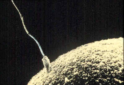
Main article: Human fertilization A sperm fertilizing an ovum
When semen is deposited in the vagina, the spermatozoa travel through the cervix and body of the uterus and into the Fallopian tubes. Fertilization of the ovum (egg cell) usually takes place in the Fallopian tube. Many sperm must cooperate to penetrate the thick protective shell-like barrier that surrounds the ovum. The first sperm that penetrates fully into the egg donates its genetic material (DNA). The egg then polarizes, repelling any additional sperm. The resulting combination is called a zygote, a new and genetically unique human organism. The term "conception" refers variably to either fertilization or to formation of the conceptus after uterine implantation, and this terminology is controversial.
Prior to fertilization, each ovum contains a complete human genome, including a single X but no Y chromosome. Likewise, each spermatozoon contains a complete set of autosomes and a single sex chromosome, either X or Y. The resulting human zygote is similar to the majority of somatic cells because it contains two copies of the genome in a diploid set of chromosomes. One set of chromosomes came from the nucleus of the ovum and the second set from the nucleus of the sperm. If the spermatozoon contributes a Y chromosome then the zygote will develop as a male. Unlike the X chromosome, the Y chromosome contains very little genetic information. However it does contain a gene, SRY, which will switch on androgen production at a later stage, leading to the development of a male body type. In contrast, the mitochondrial genetic information of the zygote comes entirely from the mother via the ovum.
Embryonic period
Main article: Human embryogenesis The initial stages of human embryogenesis
The embryonic period in humans begins at fertilization (penetration of the egg by the sperm) and continues until the end of the 10th week of gestation (8th week by embryonic age).
The embryo spends the next few days traveling down the Fallopian tube. It starts out as a single cell zygote and then divides several times to form a ball of cells called a morula. Further cellular division is accompanied by the formation of a small cavity between the cells. This stage is called a blastocyst. Up to this point there is no growth in the overall size of the embryo, as it is confined within a glycoprotein shell, known as the zona pellucida. Instead, each division produces successively smaller cells.
The blastocyst reaches the uterus at roughly the fifth day after fertilization. It is here that lysis of the zona pellucida occurs. This process is analogous to zona hatching, a term that refers to the emergence of the blastocyst from the zona pellucida, when incubated in vitro. This allows the trophectoderm cells of the blastocyst to come into contact with, and adhere to, the endometrial cells of the uterus. The trophectoderm will eventually give rise to extra-embryonic structures, such as the placenta and the membranes. The embryo becomes embedded in the endometrium in a process called implantation. In most successful pregnancies, the embryo implants 8 to 10 days after ovulation (Wilcox et al. 1999). The embryo, the extra-embryonic membranes, and the placenta are collectively referred to as a conceptus, or the "products of conception".
Rapid growth occurs and the embryo's main external features begin to take form. This process is called differentiation, which produces the varied cell types (such as blood cells, kidney cells, and nerve cells). A spontaneous abortion, or miscarriage, in the first trimester of pregnancy is usually[4] due to major genetic mistakes or abnormalities in the developing embryo. During this critical period (most of the first trimester), the developing embryo is also susceptible to toxic exposures, such as:
- Alcohol, certain drugs, and other toxins that cause birth defects, such as Fetal alcohol syndrome
- Infection (such as rubella or cytomegalovirus)
- Radiation from x-rays or radiation therapy
- Nutritional deficiencies such as lack of folate which contributes to spina bifida
Generally, if a structure pre-dates another structure in evolutionary terms, then it often appears earlier than the other in an embryo; this general observation is sometimes summarized by the phrase "ontogeny recapitulates phylogeny."[5] For example, the backbone is a common structure among all vertebrates such as fish, reptiles and mammals, and the backbone also appears as one of the earliest structures laid out in all vertebrate embryos. The cerebrum in humans, which is the most sophisticated part of the brain, develops last. The concept of recapitulation is not absolute, but it is recognized as being partly applicable to development of the human embryo.[5]
Changes by weeks of gestation
See also: Embryo and Human embryogenesisGestational age vs. embryonic age
Gestational age is the time that has passed since the onset of the last menstruation, which generally or as standard occurs 2 weeks before the actual fertilization. Embryonic age, in contrast measures the actual age of the embryo or fetus from the time of fertilization. Nevertheless, menstruation has historically been the only means of estimating embryonal/fetal age, and is still the presumed measure if not else specified. However, the actual duration between last menstruation and fertilization may in fact differ from the standard 2 weeks by several days.
Thus, the first week of embryonic age is already week three counting with gestational age.
Furthermore, the number of the week is one more than the actual age of the embryo/fetus. For example, the embryo is 0 whole weeks old during the 1st week after fertilization.
The following table summarizes the various expression systems during week number x of gestation.
Week
numberReached age
(whole weeks)Gestational x x-1 Embryonic x-2 x-3 Week 1–2
Gestational age: 0 to 1 (whole) weeks old. 1–14 days from last menstruation.
Embryonic age: -2 to -1 weeks old. (Week 1–2 of gestational age are theoretical extrapolations of embryonic age, since the fertilization hasn't actually occurred yet.)
Week 3
Gestational age: 2 (whole) weeks old. 15–21 days from last menstruation.
Embryonic age: Week nr 1. 0 (whole) weeks old. 1–7 days from fertilization.
- Fertilization of the ovum to form a new human organism, the human zygote. (day 1 of fert.[6])
- The zygote undergoes mitotic cellular divisions, but does not increase in size. This mitosis is also known as cleavage. A hollow cavity forms marking the blastocyst stage. (day 1.5–3 of fert.[6])
- The blastocyst contains only a thin rim of trophoblast cells and a clump of cells at one end known as the "embryonic pole" which include embryonic stem cells.
- The embryo hatches from its protein shell (zona pellucida) and performs implantation onto the endometrial lining of the mother's uterus. (day 5–6 of fert.[6])
- If separation into identical twins occurs, 1/3 of the time it will happen before day 5.[7]
Week 4
Gestational age: 3 weeks old. 22–28 days from last menstruation.
Embryonic age: Week nr 2. 1 week old. 8–14 days from fertilization.
- Trophoblast cells surrounding the embryonic cells proliferate and invade deeper into the uterine lining. They will eventually form the placenta and embryonic membranes. The blastocyst is fully implanted day 7–12 of fert.[6]
- Formation of the yolk sac.
- The embryonic cells flatten into a disk, two cells thick.
- If separation into identical twins occurs, 2/3 of the time it will happen between days 5 and 9. If it happens after day 9, there is a significant risk of the twins being conjoined.
- Primitive streak develops. (day 13 of fert.[6])
- Primary stem villi appear. (day 13 of fert.[6])
Week 5
Gestational age: 4 weeks old. 29–35 days from last menstruation.
Embryonic age: Week nr 3. 2 weeks old. 15–21 days from fertilization.
- A notochord forms in the center of the embryonic disk. (day 16 of fert.[6])
- Gastrulation commences. (day 16of fert.[6])
- A neural groove (future spinal cord) forms over the notochord with a brain bulge at one end. Neuromeres appear. (day 18 of fert.[6])
- Somites, the divisions of the future vertebra, form. (day 20 of fert.[6])
- Primitive heart tube is forming. Vasculature begins to develop in embryonic disc. (day 20 of fert.[6])

 A 10mm embryo from an ectopic pregnancy, still in the oviduct. This embryo is about five weeks old (or from the seventh week of menstrual age).
Week 6
Gestational age: 5 weeks old. 36–42 days from last menstruation.
Embryonic age: Week nr 4. 3 weeks old. 22–28 days from fertilization.
- The embryo measures 4 mm (1/8 inch) in length and begins to curve into a C shape.
- The heart bulges, further develops, and begins to beat in a regular rhythm. Septum primum appear.[6]
- Branchial arches, grooves which will form structures of the face and neck, form.
- The neural tube closes.
- The ears begin to form as otic pits.
- Arm buds and a tail are visible.
- Pulmonary primordium, the first traits of the lung appear.[6]
- Hepatic plate, the first traits of the liver appear.[6]
- Buccopharyngeal membrane ruptures. This is the future mouth.[6]
- Cystic diverticulum, which will become the gallbladder, and dorsal pancreatic bud, which will become the pancreas appear.[6]
- Urorectal septum begins to form. Thus, the rectal and urinary passageways become separated.[6]
- Anterior and posterior horns differentiate in the spinal cord [6]
- Spleen appears.[6]
- Ureteric buds appear.[6]
 This embryo is also from an ectopic pregnancy, this one in the cornu (the part of the uterus to which the Fallopian tube is attached). The features are consistent with a developmental age of seven weeks (reckoned as the ninth week of pregnancy).
Week 7
Gestational age: 6 weeks old. 43–49 days from last menstruation.
Embryonic age: Week nr 5. 4 weeks old. 29–35 days from fertilization.
- The embryo measures 9 mm (1/4 inch) in length.
- Lens pits and optic cups form the start of the developing eye.
- Nasal pits form.
- The brain divides into 5 vesicles, including the early telencephalon.
- Leg buds form and hands form as flat paddles on the arms.
- Rudimentary blood moves through primitive vessels connecting to the yolk sac and chorionic membranes.
- The metanephros, precursor of the definitive kidney, starts to develop.
- The initial stomach differentiation begins.
Week 8
Gestational age: 7 weeks old. 50–56 days from last menstruation.
Embryonic age: Week nr 6. 5 weeks old. 36–42 days from fertilization.
- The embryo measures 13 mm (1/2 inch) in length.
- Lungs begin to form.
- The brain continues to develop.
- Arms and legs have lengthened with foot and hand areas distinguishable.
- The hands and feet have digits, but may still be webbed.
- The gonadal ridge begins to be perceptible.
- The lymphatic system begins to develop.
- Main development of external genitalia starts.
Week 9
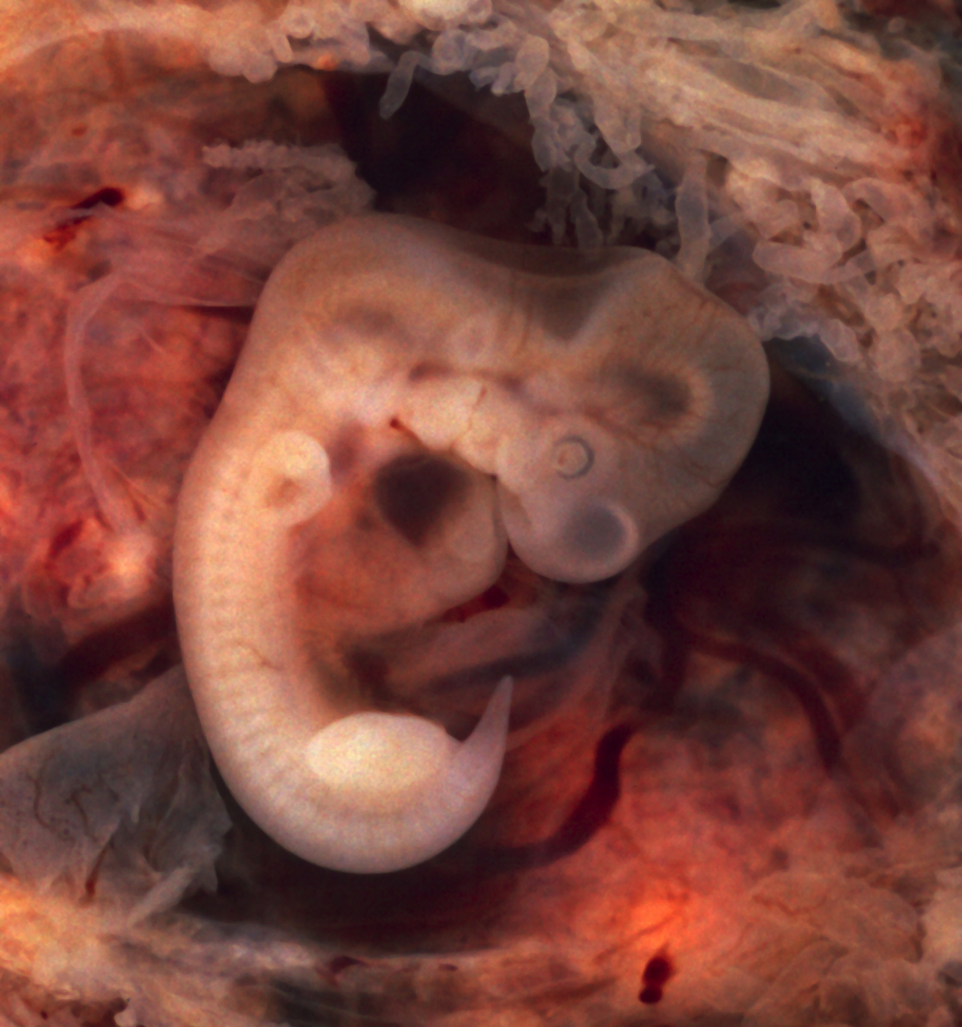
 A six week embryonic age or eight week gestational age intact human embryo.
Gestational age: 8 weeks old. 57–63 days from last menstruation.
Embryonic age: Week nr 7. 6 weeks old. 43–49 days from fertilization.
- The embryo measures 18 mm (3/4 inch) in length.
- Fetal heart tone (the sound of the heart beat) can be heard using doppler.
- Nipples and hair follicles begin to form.
- Location of the elbows and toes are visible.
- Spontaneous limb movements may be detected by ultrasound.
- All essential organs have at least begun
- The vitelline duct normally closes
Fetal period
The fetal period begins at the end of the 10th week of gestation (8th week of development). Since the precursors of all the major organs are created by this time, the fetal period is described both by organ and by a list of changes by weeks of gestational age.
Because the precursors of the organs are formed, fetus also is not as sensitive to damage from environmental exposures as the embryo. Instead, toxic exposures often cause physiological abnormalities or minor congenital malformation.
Changes by organ
Each organ has its own development.
- Development of circulatory system
- Development of digestive system
- Development of endocrine system
- Development of integumentary system
- Development of lymphatic system
- Development of muscular system
- Development of nervous system
- Development of the urinary and reproductive system
- Development of respiratory system
Changes by weeks of gestation
From the 8th week until birth (around 38 weeks), the developing organism is called a fetus. The fetus is not as sensitive to damage from environmental exposures as the embryo, and toxic exposures often cause physiological abnormalities or minor congenital malformation. All major structures are already formed in the fetus, but they continue to grow and develop.
 Fetus at 8 weeks after fertilization.[10]
Weeks 10–12
Gestational age: 9–11 weeks old.
Embryonic age: Weeks nr 8–10. 7–9 weeks old.
- Embryo measures 30–80 mm (1.2–3.2 inches) in length.
- Ventral and dorsal pancreatic buds fuse during the 8th week
- Intestines rotate.
- Facial features continue to develop.
- The eyelids are more developed.
- The external features of the ear begin to take their final shape.
- The head comprises nearly half of the fetus' size.
- The face is well formed
- The eyelids close and will not reopen until about the 28th week.
- Tooth buds, which will form the baby teeth, appear.
- The limbs are long and thin.
- The fetus can make a fist with its fingers.
- Genitals appear well differentiated.
- Red blood cells are produced in the liver.
Weeks 13 to 16
Gestational age: 12–15 weeks old.
Embryonic age: Weeks nr 11–14. 10–13 weeks old.
- The fetus reaches a length of about 15 cm (6 inches).
- A fine hair called lanugo develops on the head.
- Fetal skin is almost transparent.
- More muscle tissue and bones have developed, and the bones become harder.
- The fetus makes active movements.
- Sucking motions are made with the mouth.
- Meconium is made in the intestinal tract.
- The liver and pancreas produce fluid secretions.
- From week 13, sex prediction by obstetric ultrasonography is almost 100% accurate.[11]
- At week 15, main development of external genitalia is finished
 Fetus at 18 weeks after fertilization.[12]
Week 20
Gestational age: 18 weeks old.
Embryonic age: Week nr 17. 16 weeks old.
- The fetus reaches a length of 20 cm (8 inches).
- Lanugo covers the entire body.
- Eyebrows and eyelashes appear.
- Nails appear on fingers and toes.
- The fetus is more active with increased muscle development.
- "Quickening" usually occurs (the mother and others can feel the fetus moving).
- The fetal heartbeat can be heard with a stethoscope.
Week 23
Gestational age: 22 weeks old.
Embryonic age: Week nr 21. 20 weeks old.
- The fetus reaches a length of 28 cm (11.2 inches).
- The fetus weighs about 925g.
- Eyebrows and eyelashes are well formed.
- All of the eye components are developed.
- The fetus has a hand and startle reflex.
- Footprints and fingerprints continue forming.
- Alveoli (air sacs) are forming in lungs.
Week 27
Gestational age: 26 weeks old.
Embryonic age: Week nr 25. 24 weeks old.
- The fetus reaches a length of 38 cm (15 inches).
- The fetus weighs about 1.2 kg (2 lb 11 oz).
- The brain develops rapidly.
- The nervous system develops enough to control some body functions.
- The eyelids open and close.
- The cochleae are now developed, though the myelin sheaths in neural portion of the auditory system will continue to develop until 18 months after birth.
- The respiratory system, while immature, has developed to the point where gas exchange is possible.
Week 31
Gestational age: 30 weeks old.
Embryonic age: Week nr 29. 28 weeks old.
- The fetus reaches a length of about 38–43 cm (15–17 inches).
- The fetus weighs about 1.5 kg (3 lb 0 oz).
- The amount of body fat rapidly increases.
- Rhythmic breathing movements occur, but lungs are not fully mature.
- Thalamic brain connections, which mediate sensory input, form.
- Bones are fully developed, but are still soft and pliable.
- The fetus begins storing a lot of iron, calcium and phosphorus.
Week 35
Gestational age: 34 weeks old.
Embryonic age: Week nr 33. 32 weeks old.
- The fetus reaches a length of about 40–48 cm (16–19 inches).
- The fetus weighs about 2.5 to 3 kg (5 lb 12 oz to 6 lb 12 oz).
- Lanugo begins to disappear.
- Body fat increases.
- Fingernails reach the end of the fingertips.
- A baby born at 36 weeks has a high chance of survival, but may require medical interventions.
 Fetus at 38 weeks after fertilization.[13]
Weeks 36 to 39
Gestational age: 35–38 weeks old.
Embryonic age: Weeks nr 34–37. 33–36 weeks old.
- The fetus is considered full-term at the end of the 37th week of gestational age.
- It may be 48 to 53 cm (19 to 21 inches) in length.
- The lanugo is gone except on the upper arms and shoulders.
- Fingernails extend beyond fingertips.
- Small breast buds are present on both sexes.
- Head hair is now coarse and thickest.
The development is continued postnatally with adaptation to extrauterine life and child development stages.
Growth rate
The growth rate of an embryo and infant can be reflected as the weight per gestational age, and is often given as the weight put in relation to what would be expected by the gestational age. A baby born within the normal range of weight for that gestational age is known as appropriate for gestational age (AGA). An abnormally slow growth rate results in the infant being small for gestational age, and, on the other hand, an abnormally large growth rate results in the infant being large for gestational age. A slow growth rate and preterm birth are the two factors that can cause a low birth weight.
See also
References
- ^ Definitions and Indicators in Family Planning. Maternal & Child Health and Reproductive Health. By European Regional Office, World Health Organization. Revised March 1999 & January 2001. In turn citing: WHO Geneva, WHA20.19, WHA43.27, Article 23
- ^ patient.co.uk » PatientPlus » Antepartum Haemorrhage Last Updated: 5 May 2009
- ^ The Royal Women’s Hospital > antepartum haemorrhage Retrieved on Jan 13, 2009
- ^ Moore L. Keith. (2008). Before We Are Born: Essentials of Embryology and Birth Defects. Philadelphia, PA: Saunders/Elsevier. ISBN 978-1-4160-3705-7.
- ^ a b Stephen Jay Gould,. Ontogeny and Phylogeny. Cambridge, Mass: Belknap Press. p. 206. ISBN 0-674-63941-3. http://www.sjgarchive.org/library/ontogeny.html.
- ^ a b c d e f g h i j k l m n o p q r s t )William J. Larsen (2001). Human embryology. Edinburgh: Churchill Livingstone. ISBN 0-443-06583-7.[page needed]
- ^ Scott F. Gilbert; with a chapter on plant development by Susan R. Singer (2000). Developmental biology. Sunderland, Mass: Sinauer Associates. ISBN 0-87893-243-7. http://www.ncbi.nlm.nih.gov/books/bv.fcgi?rid=dbio.box.2669.
- ^ 3D Pregnancy (large image of fetus at 4 weeks after fertilization). Retrieved 2007-08-28. A rotatable 3D version of this photo is available here, and a sketch is available here.
- ^ Wagner F, Erdösová B, Kylarová D (December 2004). "Degradation phase of apoptosis during the early stages of human metanephros development". Biomed Pap Med Fac Univ Palacky Olomouc Czech Repub 148 (2): 255–6. PMID 15744391.
- ^ 3D Pregnancy (large image of fetus at 10 weeks after fertilization). Retrieved 2007-08-28. A rotatable 3D version of this photo is available here, and a sketch is available here.
- ^ Mazza V, Falcinelli C, Paganelli S, et al. (June 2001). "Sonographic early fetal gender assignment: a longitudinal study in pregnancies after in vitro fertilization". Ultrasound Obstet Gynecol 17 (6): 513–6. doi:10.1046/j.1469-0705.2001.00421.x. PMID 11422974.
- ^ 3D Pregnancy (large image of fetus at 18 weeks after fertilization). Retrieved 2007-08-28. A rotatable 3D version of this photo is available here, and a sketch is available here.
- ^ 3D Pregnancy (large image of fetus at 38 weeks after fertilization). Retrieved 2007-08-28. A rotatable 3D version of this photo is available here, and a sketch is available here.
- MedlinePlus Encyclopedia Fetal development
- Moore, Keith L.. The Developing Human (3rd ed.). Philadelphia PA: W.B. Saunders Company.
- Wilcox AJ, Baird DD, Weinberg CR (June 1999). "Time of implantation of the conceptus and loss of pregnancy". N. Engl. J. Med. 340 (23): 1796–9. doi:10.1056/NEJM199906103402304. PMID 10362823. http://www.nejm.org/doi/abs/10.1056/NEJM199906103402304?url_ver=Z39.88-2003&rfr_id=ori:rid:crossref.org&rfr_dat=cr_pub%3dpubmed.
- Ljunger E, Cnattingius S, Lundin C, Annerén G (November 2005). "Chromosomal anomalies in first-trimester miscarriages". Acta Obstet Gynecol Scand 84 (11): 1103–7. doi:10.1111/j.0001-6349.2005.00882.x. PMID 16232180. http://onlinelibrary.wiley.com/resolve/openurl?genre=article&sid=nlm:pubmed&issn=0001-6349&date=2005&volume=84&issue=11&spage=1103.
External links
- Fetal Development Timeline from AboutKidsHealth.ca
- The Changes in Each Stage of Human Development
- Real Time Presentation of Human Embryo Development
- Fetal development week by week at www.babycentre.co.uk
- Pregnancy Week by Week
Human Development: Biological • Psychological Pre- and perinatal BiologicalPrenatal developmentPsychologicalInfancy BiologicalPsychologicalInfant and child psychologyChildhood BiologicalPsychologicalInfant and child psychology · PreadolescenceAdolescence BiologicalPsychologicalYoung adulthood PsychologicalMiddle adulthood BiologicalMaturity BiologicalPsychologicalLegal and general definitions Theorists and theories Human physiology and endocrinology of sexual reproduction Menstrual and estrous cycle Gametogenesis Spermatogenesis (spermatogonium, spermatocyte, spermatid, sperm) · Oogenesis (oogonium, oocyte, ootid, ovum) · Germ cell (gonocyte, gamete)Human sexual behavior Sexual intercourse · Masturbation · Erection · Orgasm · Ejaculation · Insemination · Fertilisation/Fertility · Implantation · Pregnancy · Postpartum period · Mechanics of sexLife span Prenatal development/Sexual dimorphism/Sexual differentiation (Feminization, Virilization) · Puberty (Gonadarche, Pubarche, Menarche, Adrenarche) ·
Maternal age / Paternal age · Climacteric (Menopause, Andropause)Egg (biology) Reproductive endocrinology
and infertilityBreast Pregnancy and childbirth Planning Conception Testing Prenatal AnatomyDevelopmentProceduresChildbirth PreparationRolesDeliveryPostpartum Obstetric history Developmental biology > Human embryogenesis (development of embryo) and development of fetus (TE E2.0) First three
weeksWeek 1Fertilization · Oocyte activation · Zygote · Cleavage · Morula · Blastula (Blastomere) · Blastocyst · Inner cell massWeek 2
(Bilaminar)Week 3
(Trilaminar)Archenteron/Primitive streak (Primitive pit, Primitive knot/Blastopore, Primitive groove) · Gastrula/Gastrulation · Regional specification · Embryonic discSplanchnopleuric mesenchymeChorda- · Paraxial (Somite/Somitomere) · Intermediate · Lateral plate (Intraembryonic coelom, Splanchnopleuric mesenchyme/Somatopleuric mesenchyme)Categories:


Wikimedia Foundation. 2010.