- Cryo-electron microscopy
-
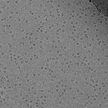 CryoEM image of GroEL suspended in vitreous ice at 50,000X magnification.
Cryo-electron microscopy (cryo-EM), or electron cryomicroscopy, is a form of transmission electron microscopy (EM) where the sample is studied at cryogenic temperatures (generally liquid nitrogen temperatures). Cryo-EM is developing popularity in structural biology.
The popularity of cryoelectron microscopy stems from the fact that it allows the observation of specimens that have not been stained or fixed in any way, showing them in their native environment, in contrast to X-ray crystallography, which generally requires placing the samples in non-physiological environments, which can occasionally lead to functionally irrelevant conformational changes. In practice, the resolution of cryo-electron microscopy maps is not high enough to allow for unambiguous model construction on the basis of EM maps only, and models obtained by protein crystallography are used to interpret the cryo-EM maps. However, the resolution of cryo-EM maps is improving steadily, and some virus structures obtained by cryo-EM are already at a resolution that can be interpreted in terms of an atomic model.
A version of electron cryomicroscopy is cryo-electron tomography (CET) where a 3D reconstruction of a sample is created from tilted 2D images.
Contents
Development
The original rationale for cryoelectron microscopy was as a means to fight radiation damage for biological specimens. The amount of radiation required to collect an image of a specimen in the electron microscope is comparable to placing the sample about 20 m away from a thermonuclear device[citation needed]. In addition, the high vacuum required on the column of an electron microscope makes the environment for the sample quite harsh.
The problem of the vacuum was partially solved by the introduction of negative stains such as uranium salts (uranyl acetate is perhaps the most common negative stain), but even with negative stains, biological samples are prone to structural collapse upon dehydration of the specimen. Embedding the samples in ice below the sublimation temperature was a possibility that was contemplated early on, but water tends to arrange into a crystalline lattice of lower density upon freezing and this tends to destroy the structure of anything that is embedded in it.
In the early 80's, several groups studying solid state physics were attempting to produce vitreous ice by different means, such as high pressure freezing or flash freezing. Theoretical calculations deemed this task impossible, but in a seminal paper in 1984, the group led by Jacques Dubochet at the European Molecular Biology Laboratory showed images of adenovirus embedded in a vitrified layer of water[citation needed]. This paper is generally considered to mark the birth of cryoelectron microscopy, and the technique has been developed to the point of becoming routine at several laboratories throughout the world.
Biological specimens
Thin film
The biological material is spread on an electron microscopy grid and is preserved in a frozen-hydrated state by rapid freezing, usually in liquid ethane near liquid nitrogen temperature. By maintaining specimens at liquid nitrogen temperature or colder, they can be introduced into the high-vacuum of the electron microscope column. Most biological specimens are extremely radiation sensitive, so they must be imaged with low-dose techniques (usefully, the low temperature of cryo-electron microscopy provides an additional protective factor against radiation damage).
Consequently, the images are extremely noisy. For some biological systems it is possible to average images to increase the signal-to-noise ratio and retrieve high-resolution information about the specimen using the technique known as single particle analysis. This approach in general requires that the things being averaged are identical, although some limited conformational heterogeneity can now be studied (e.g. ribosome). Three dimensional reconstructions from cryo-EM images of protein complexes and viruses have been solved to sub-nanometer or near-atomic resolution, allowing new insights into the structure and biology of these large assemblies.
Analysis of ordered arrays of protein, such as 2-D crystals of transmembrane proteins or helical arrays of proteins, also allows a kind of averaging which can provide high-resolution information about the specimen. This technique is called electron crystallography.
Vitreous sections
The thin film method is limited to thin specimens (typically < 500 nm) because the electrons cannot cross thicker samples without multiple scattering events. Thicker specimens can be vitrified by plunge freezing (cryofixation) in ethane (up to tens of μm in thickness) or more commonly by high pressure freezing (up to hundreds of μm). They can then be cut in thin sections (40 to 200 nm thick) with a diamond knife in a cryoultramicrotome at temperatures lower than -135 °C (devitrification temperature). The sections are collected on an electron microscope grid and are imaged in the same manner as specimen vitrified in thin film. This technique is called cryo-electron microscopy of vitreous sections (CEMOVIS) or cryo-electron microscopy of frozen-hydrated sections.
Techniques in cryoelectron microscopy
Cryoelectron microscopy has a variety of techniques which can be used.[1] Three most popular ones are:
- Single particle analysis
- Cryo-electron tomography
- Freeze trapping
References
See also
- Software tools for molecular microscopy
- Single particle analysis
- Resolution (electron density)
- EM Data Bank
Further reading
- Frank, Joachim (2006). Three-Dimensional Electron Microscopy of Macromolecular Assemblies. New York: Oxford University Press. ISBN 0-19-518218-9.
- van Heel M, Gowen B, Matadeen R, Orlova EV, Finn R, Pape T, Cohen D, Stark H, Schmidt R, Schatz M, Patwardhan A (2000). "Single-particle electron cryo-microscopy: towards atomic resolution.". Q Rev Biophys. 33: 307–69.
External links
- "EM for Dummies". EM for Dummies. http://cryoem.berkeley.edu/~nieder/em_for_dummies/. Retrieved June 9, 2006.
- The Fine Structure of a Frozen Virus - Sophisticated single-particle electron cryomicroscopy reveals unprecedented details in a virus's protein coat, Technology Review, March 19, 2008
Primary database:
Protein structural analysis High resolution Medium resolution Fiber diffraction | Mass spectrometry | SAXSSpectroscopic Translational Diffusion Rotational Diffusion Chemical Thermodynamic Computational ←Tertiary structureCategories:- Microscopes
- Cell biology
- Protein structure

Wikimedia Foundation. 2010.