- Facial suture (trilobite anatomy)
-
Facial sutures are sutures in the cephalon (head) of most trilobites, along which the exoskeleton splits during molting.
Contents
Description
Facial or Cephalic sutures are the natural fracture lines in the cephalon of trilobites. Their function is to assist the trilobite in shedding its old exoskeleton during ecdysis (or molting).[1]
Primitive trilobites from the Early Cambrian (like Fallotaspis, Eofallotaspis, Schmidtiellus, and Profallotaspis) lacked facial sutures. They are all classified under the suborder Olenellina. They are believed to have never developed facial sutures, having pre-dated their evolution. Because of this (along with other primitive characteristics), they are thought to be the earliest ancestors of later trilobites.[2][3]
Some other later trilobites also lost facial sutures secondarily.[2] The type of sutures found in different species are used extensively in the taxonomy and phylogeny of trilobites.[4]
Dorsal sutures
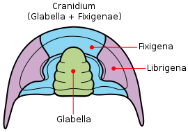
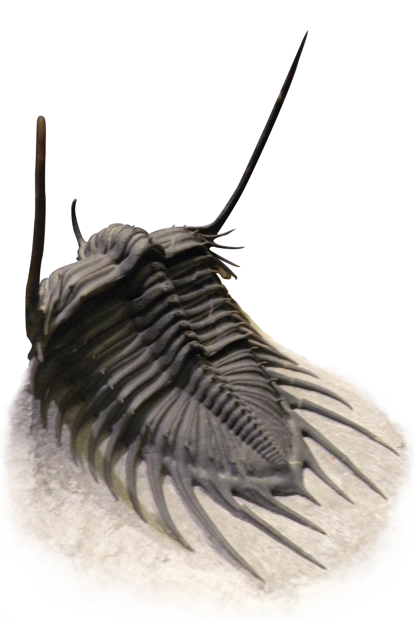
The dorsal surface of the trilobite cephalon (the frontmost tagma, or the 'head') can be divided into two regions - the cranidium and the librigena ("free cheeks"). The cranidium can be further divided into the glabella (the central lobe in the cephalon) and the fixigena ("fixed cheeks").[5] The facial sutures lie along the anterior edge, at the division between the cranidium and the librigena.
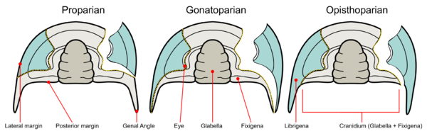
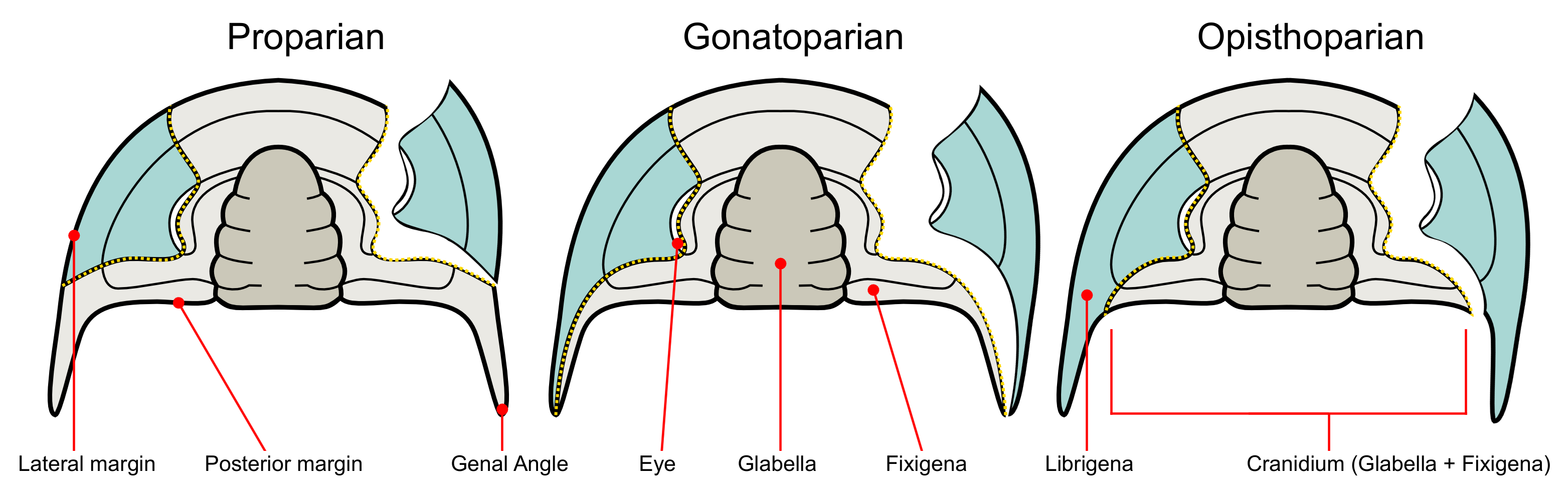
Trilobite facial sutures on the dorsal side can be roughly divided into three main types according to where the sutures end relative to the genal angle (the edges where the side and rear margins of the cephalon converge).[6]
- Proparian - The facial suture ends ahead of the genal angle, along the lateral margin.[5] Example genera showing this type of suture include Dalmanites of Phacopina (Phacopida) and Ekwipagetia of Eodiscina (Agnostida).
- Gonatoparian - The facial suture ends at the tip of the genal angle. It is also known as Marginal or Hypoparian.[7] Example genera showing this type of suture include Calymene and Trimerus of Calymenina (Phacopida).[4]
- Opisthoparian - The facial suture ends at the posterior margin of the cephalon.[4] Example genera showing this type of suture include Peltura of Olenina (Ptychopariida) and Bumastus of Illaenina (Corynexochida). This is the most common type of facial suture.[4]
In some trilobites the sutures may be difficult to see as they run along the margins of the cephalon.[5] This is considered a fourth type of suture known as Marginal (or Hypoparian sutures). In this type, the sutures run mostly or wholly along the margin of the cephalon. They are not considered to be a formal phylogenetic grouping like the previous groups as they have independently developed in several groups of trilobites. Other types of sutures became more marginal when trilobite groups acquire secondary blindnesss (a “devolution” of visual organs in some trilobites).[8] The marginal sutures exhibited by the harpetids and trinucleioids, for example, are derived from opisthoparian sutures.[9] Example genera from both groups are Harpes and Cryptolithus, both of which are blind.[4]
There are also two types of sutures in the dorsal surface connected to the compound eye.[8][4] They are:
- Ocular suture - are sutures surrounding the edges of the compound eye. Trilobites with these sutures lose the entrie surface of the eyes when molting. It is common among Cambrian trilobites.
- Palepebral suture - are sutures which form part of the dorsal facial suture running along the top edges of the compound eye.
Ventral sutures
Dorsal facial sutures continue downward to the ventral side of the cephalon where they become the Connective sutures that divide the doublure (the parts of the exoskeleton that extends into the ventral side of the trilobite). The following are the types of ventral sutures.[8]
- Connective sutures - are the sutures that continue from the facial sutures past the front margin of the cephalon.
- Rostral suture - is only present when the trilobite possesses a rostrum (or rostral plate). It connects the rostrum to the front part of the dorsal cranidium.
- Hypostomal suture - separates the hypostome from the doublure when the hypostome is of the attached type. It is absent when the hypostome is free-floating (i.e. natant). it is also absent in some coterminant hpostomes where the hypostome is fused to the doublure.
- Median suture - exhibited by asaphid trilobites, they are formed when instead of becoming connective sutures, the two dorsal sutures converge at a point in front of the cephalon then divide straight down the center of the doublure.
See also
References
- ^ Riccardo Levi-Setti (1995). Trilobites. University of Chicago Press. ISBN 9780226474526. http://books.google.com/books?id=rRyHMz-FkJUC&printsec=frontcover.
- ^ a b Chris Clowes (April 15, 2006). "Trilobite Origins". Peripatus. http://www.peripatus.gen.nz/taxa/arthropoda/trilobita/TriOri.html. Retrieved April 13, 2011.
- ^ B. S., Lieberman (2002), "Phylogenetic analysis of some basal early Cambrian trilobites, the biogeographic origins of the eutrilobita, and the timing of the Cambrian radiation", Journal of Paleontology 76: 692–708, doi:10.1666/0022-3360(2002)076<0692:PAOSBE>2.0.CO;2
- ^ a b c d e f Samuel M. Gon III (February 3, 2009). "Trilobite Facial Sutures". A Guide to the Orders of Trilobites. http://www.trilobites.info/sutures.htm. Retrieved April 13, 2011.
- ^ a b c Rhona M. Black (1988), The elements of palaeontology (2 ed.), Cambridge University Press, pp. 151–152, ISBN 9780521348362, http://books.google.com/books?id=cEh5Yn0neiUC&printsec=frontcover
- ^ Michael Kipping. "Change of suit". www.trilobita.de. http://www.trilobita.de/english/molting.htm. Retrieved April 13, 2011.
- ^ Pat Vickers Rich, Mildred Adams Fenton, Carroll Lane Fenton, Thomas Hewitt Rich (1989). The fossil book: a record of prehistoric life. Dover books on animals. Courier Dover Publications. p. 204. ISBN 9780486293714. http://books.google.com/books?id=_ntSspji0LYC&printsec=frontcover.
- ^ a b c Euan Neilson Kerr Clarkson (1998). Invertebrate palaeontology and evolution. Wiley-Blackwell. ISBN 9780632052387. http://books.google.com/books?id=g1P2VaPQWfUC&printsec=frontcover#v=onepage&q&f=false.
- ^ Euan Clarkson, Riccardo Levi-Setti, and Gabor Horvath (2006). "The eyes of trilobites: The oldest preserved visual system". Arthropod Structure & Development (Elsevier) 35: 247–259. doi:10.1016/j.asd.2006.08.002.
External links
Trilobites 
Taxonomy Geochronology Cambrian · Ordovician · Silurian · Devonian · Carboniferous · Permian
Notable fossils Acropyge · Calymene · Cheiropyge · Eofallotaspis tioutensis · Hupetina antiqua · Isotelus · Kathwaia · Misszhouia · Olenus · Ogygiocarella debuchii · Paraphillipsia · Phacops rana · Profallotaspis jakutensis · Pseudophillipsia · Serrania gordaensis
Notable trilobitologists Alexander von Humboldt · Joachim Barrande · Alfred Russel Wallace · Charles Doolittle Walcott · Charles Emerson Beecher · Roderick Murchison · Adam Sedgwick · Rudolf Kaufmann · Rudolf Richter & Emma Richter · Martin Basse · Niles Eldredge · George Frederic Matthew · Harry B. Whittington · Brian D. E. Chatterton · Richard Fortey · Euan Clarkson · Franco Rasetti · Gregory D. Edgecombe · Bruce S. Lieberman · Gerald J. Kloc · Gerhard K. B. Alberti · Teiichi Kobayashi
Fossil sites Anti-Atlas Mountains · Beecher's Trilobite Bed · Burgess Shale · Emu Bay shale · Gees, Gerolstein · Hunsrück Slate · Jince Formation · Latham Shale · Llandeilo Group · Maotianshan Shales · Silica Shale Formation · Walcott-Rust quarry · Wenlock Group · Wheeler Shale · Wolchov River · Wren's NestMorphology Cranidium · Effacement · Enrollment · Facial sutures · Fixigena · Gena · Genal angle · Glabella · Gonatoparian · Heteronomous · Homonomous · Hypostome · Isopygous · Librigena · Macropygous · Micropygous · Opisthoparian · Proparian · Prosopon · Spinosity · SubisopygousCategory:TrilobitesCategories:- Trilobites
- Arthropod anatomy

Wikimedia Foundation. 2010.