- Midbrain
-
Brain: Mesencephalon 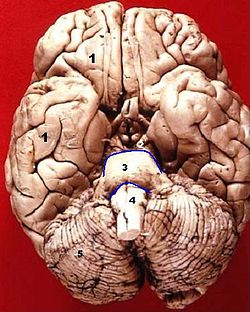
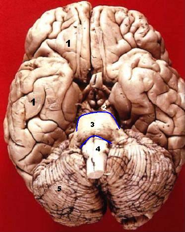
Inferior view mesencephalon (2), above (3) 
Human brainstem mesencephalon (B) Latin mesencephalon Gray's subject #188 800 NeuroNames hier-445 MeSH Mesencephalon The midbrain or mesencephalon (from the Greek mesos - middle, and enkephalos - brain[1]) is a portion of the central nervous system associated with vision, hearing, motor control, sleep/wake, arousal (alertness), and temperature regulation.[2]
Anatomically, it comprises the tectum (or corpora quadrigemina), tegmentum, the ventricular mesocoelia (or "iter"), and the cerebral peduncles, as well as several nuclei and fasciculi. Caudally the mesencephalon adjoins the pons (metencephalon) and rostrally it adjoins the diencephalon (Thalamus, hypothalamus, etc.). The midbrain is located below the cerebral cortex, and above the hindbrain placing it near the center of the brain.[3]
Contents
Development
During embryonic development, the midbrain arises from the second vesicle, also known as the mesencephalon, of the neural tube. Unlike the other two vesicles, the prosencephalon and rhombencephalon, the mesencephalon remains undivided for the remainder of neural development. It does not split into other brain areas. while the prosencephalon, for example, divides into the telencephalon and the diencephalon.[4]
Throughout embryonic development, the cells within the midbrain continually multiply and compress the still-forming Aqueduct of Sylvius or cerebral aqueduct. Partial or total obstruction of the cerebral aqueduct during development can lead to congenital hydrocephalus.[5]
Functional Role
The mesencephalon is considered part of the brainstem. Its substantia nigra is closely associated with motor system pathways of the basal ganglia. The human mesencephalon is archipallian in origin, meaning its general architecture is shared with the most ancient of vertebrates. Dopamine produced in the substantia nigra plays a role in motivation and habituation of species from humans to the most elementary animals such as insects.
Corpora quadrigemina
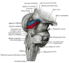
The corpora quadrigemina ("quadruplet bodies") are four solid optic lobes on the dorsal side of cerebral aqueduct, where the superior posterior pair are called the superior colliculi and the inferior posterior pair are called the inferior colliculi. The four solid optic lobes help to decussate several fibres of the optic nerve. However some fibers also show ipsilateral arrangement (i.e. they run parallel on the same side without decussating.) The superior colliculus is involved with saccadic eye movements; while the inferior is a synapsing point for sound information. The trochlear nerve comes out of the posterior surface of the midbrain, below the inferior colliculus.
Cerebral peduncle
The cerebral peduncles are paired structures, present on the ventral side of cerebral aqueduct, and they further carry tegmentum on the dorsal side and cresta or pes on the ventral side, and both of them accommodate the corticospinal tract fibres, from the internal capsule (i.e. ascending + descending tracts = longitudinal tract.) the middle part of cerebral peduncles carry substantia nigra (also called "Black Matter") which is a type of basal nucleus. It is the only part of the brain that carries melanin pigment.
Between the peduncles is the interpeduncular fossa, which is a cistern filled with cerebrospinal fluid. The oculomotor nerve comes out between the peduncles, and the trochlear nerve is visible wrapping around the outside of the peduncles. The oculomotor is responsible for pupil constriction (parasympathetic) and eye movement.[dubious ]
Anatomical features of cross-sections through the midbrain

The midbrain is usually sectioned at the level of the superior and inferior colliculi.
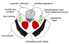
- A transverse cross-section at the level of the superior colliculus shows the red nucleus, the nuclei of the oculomotor nerve (and associated Edinger-Westphal nucleus), the superior cerebellar peduncles or crus cerebri, and the substantia nigra.[6]
- A horizontal cross section at the level of the inferior colluiculus still shows the substantia nigra. Also apparent are the trochlear nerve nucleus, and the decussation of the superior cerebellar peduncles.[7]
- Both sections will show the cerebral aqueduct, which connects the third and fourth ventricle and the periaqueductal gray.[8]
One mnemonic for remembering the structures of the midbrain involves visualizing the mesencephalic cross-section as an upside down bear face. The two red nuclei are the eyes of the bear and the cerebellar peduncles are the ears.
Organization
- mesencephalon
- tectum
- inferior colliculi
- superior colliculi
- cerebral peduncle
- midbrain tegmentum
- crus cerebri
- substantia nigra
- tectum
See also
References
- ^ Mosby’s Medical, Nursing and Allied Health Dictionary, Fourth Edition, Mosby-Year Book 1994, p. 981
- ^ Breedlove, Watson, & Rosenzweig. Biological Psychology, 6th Edition, 2010, pp. 45-46
- ^ http://www.morris.umn.edu/~ratliffj/images/brain_slides/slide_5.htm
- ^ Martin. Neuroanatomy Text and Atlas, Second Edition, 1996, pp. 35-36.
- ^ "Hydrocephalus Fact Sheet". National Institute of Neurological Disorders and Stroke. 2008-02. http://www.ninds.nih.gov/disorders/hydrocephalus/detail_hydrocephalus.htm. Retrieved 2011-03-23.
- ^ Martin. Neuroanatomy Text and Atlas, Second edition. 1996, pp. 522-525.
- ^ Martin. Neuroanatomy Text and Atlas, Second edition. 1996, pp. 522-525.
- ^ Martin. Neuroanatomy Text and Atlas, Second edition. 1996, pp. 522-525.
Nervous system (TA A14, GA 9) Central nervous system Brain: Rhombencephalon (Medulla oblongata, Pons, Cerebellum) • Mesencephalon • Prosencephalon (Diencephalon, Telencephalon)Peripheral nervous system Human brain: mesencephalon (midbrain) (TA A14.1.06, GA 9.800) Tectum
(Dorsal)SurfaceWhite: Sensory/ascendingWhite: Motor/descendingPeduncle
(Ventral)White: Sensory/ascendinglemnisci (Medial, Lateral) · Ascending MLF (Vestibulo-oculomotor fibers) · Spinothalamic tract · Anterior trigeminothalamic tract · Dentatothalamic tractWhite: Motor/descendingGrey: otherPeriaqueductal gray/Raphe nuclei (Dorsal raphe nucleus)
Ventral tegmental area • Pedunculopontine nucleus • Red nucleus
riMLFBaseWhite: Motor/descendingSurfaceCategories:


Wikimedia Foundation. 2010.