Midline shift

Midline shift
Midline shift (arrow) is present in this brain after a stroke (infarct depicted in shaded area).
This subdural hematoma/epidural hematoma (arrows) is causing midline shift of the brain

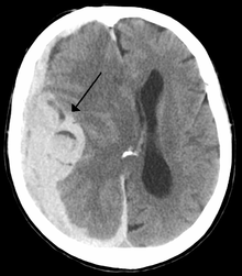
Midline shift is a shift of the brain past its center line.[1] The sign may be evident on neuroimaging such as CT scanning.[1] The sign is considered ominous because it is commonly associated with a distortion of the brain stem that can cause serious dysfunction evidenced by abnormal posturing and failure of the pupils to constrict in response to light.[1] Midline shift is often associated with high intracranial pressure (ICP), which can be deadly.[1] In fact, midline shift is a measure of ICP; presence of the former is an indication of the latter.[2] Presence of midline shift is an indication for neurosurgeons to take measures to monitor and control ICP.[1] Immediate surgery may be indicated when there is a midline shift of over 5 mm.[3] The sign can be caused by conditions including traumatic brain injury[1] and stroke that raise intracranial pressure.

See also

References

  1. ^ a b c d e f Gruen P (May 2002). "Surgical management of head trauma". Neuroimaging clinics of North America 12 (2): 339–43. doi:10.1016/S1052-5149(02)00013-8. PMID 12391640. 
  2. ^ Maas AI, Stocchetti N, Bullock R (August 2008). "Moderate and severe traumatic brain injury in adults". Lancet Neurology 7 (8): 728–41. doi:10.1016/S1474-4422(08)70164-9. PMID 18635021. 
  3. ^ Valadka AB (2004). "Injury to the cranium". In Moore EJ, Feliciano DV, Mattox KL. Trauma. New York: McGraw-Hill, Medical Pub. Division. pp. 389. ISBN 0-07-137069-2. http://books.google.com/?id=VgizxQg-8QQC&pg=PA545&dq=tracheobronchial. Retrieved 2008-08-15. 


Wikimedia Foundation. 2010.

Игры ⚽ Нужен реферат?

Look at other dictionaries:

  • Intracranial pressure — (ICP) is the pressure inside the skull and thus in the brain tissue and cerebrospinal fluid (CSF). The body has various mechanisms by which it keeps the ICP stable, with CSF pressures varying by about 1 mmHg in normal adults through shifts in… …   Wikipedia

  • Epidural hematoma — DiseaseDisorder infobox Name = Extradural haemorrhage Caption = Nontraumatic epidural hematoma in a young woman. The grey area in the top left is organizing hematoma, causing midline shift and compression of the ventricle. ICD10 =… …   Wikipedia

  • Traumatic brain injury — See also: Brain injury (disambiguation) Traumatic brain injury Classification and external resources …   Wikipedia

  • Brain tumor — Classification and external resources Brain metastasis in the right cerebral hemisphere from lung cancer shown on T1 weighted magnetic resonance imaging with intravenous contrast …   Wikipedia

  • Mass effect (medicine) — This article is about the phenomenon in medicine. For other uses, see Mass effect (disambiguation). In medicine, a mass effect is the effect of a growing mass (or tumor in its narrow meaning), for example the consequences of a growing cancer. For …   Wikipedia

  • Intrazerebrale Blutung — CT Aufnahme einer intrazerebralen Blutung (helle, zentrale Verschattungen) …   Deutsch Wikipedia

  • Death of Brian Rossiter — Brian Rossiter Died 14 September 2002 (aged 14) Cork, Ireland Cause of death blunt force trauma to the head; open verdict Bo …   Wikipedia

  • eye, human — ▪ anatomy Introduction  specialized sense organ capable of receiving visual images, which are then carried to the brain. Anatomy of the visual apparatus Structures auxiliary to the eye The orbit       The eye is protected from mechanical injury… …   Universalium

  • Hemispatial neglect — Classification and external resources Hemispatial neglect is most frequently associated with a lesion of the right parietal lobe (in yellow, at top) ICD 9 …   Wikipedia

  • Rhinoplasty — For the album by Primus, see Rhinoplasty (album). Rhinoplasty Intervention Rhinoplasty: The lower lateral cartilage (greater alar cartilage) exposed for plastic modification via the left nostril …   Wikipedia

Share the article and excerpts

Direct link
Do a right-click on the link above
and select “Copy Link”