- Nucleus proprius of spinal cord
-
Nucleus proprius of spinal cord 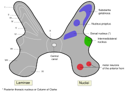
Medulla spinalis - Substantia grisea Latin nucleus proprius medullae spinalis; laminae spinales III et IV The Nucleus proprius is a layer of the spinal cord adjacent to the substantia gelatinosa. Nucleus proprius constitutes the bulk of the dorsal horn and receives inputs from the dorsal root ganglions that carry sensory information, such as light touch, as well as pain and temperature information. Cells in this nucleus project to deeper laminae of the spinal cord, to the posterior column nuclei, and to other supraspinal relay centers including the midbrain, thalamus, and hypothalamus. Rexed laminae III, IV, and V make up the nucleus proprius.[1] Nucleus proprius (NP), along with nucleus dorsalis (ND) are involved in sensing fine touch and proprioception.
See also
References
External links
- "Nucleus proprius" at Dorland's Medical Dictionary
- Diagram at pixelatedbrain.com
- Sławomirski J, Głuszak J (1986). "Structure and topography of the nucleus proprius cornus dorsalis of the spinal cord of horses". Pol Arch Weter 25 (4): 131–6. PMID 3620378.
Anatomy of torso (primarily): the spinal cord (TA 14.1.02, GA 9.749) External, dorsal Posterior median sulcus · Posterolateral sulcusGrey matter/
Rexed laminaeI–VI: Posterior hornI: Marginal nucleus · II: Substantia gelatinosa of Rolando · III+IV: Nucleus proprius · Spinal lamina V · Spinal lamina VIVII: Lateral hornVIII–IX: Anterior hornX: OtherWhite matter somatic/
ascending
(blue)Posterior/PCML: touch: Gracile · Cuneate
Lateral: proprioception: Spinocerebellar (Dorsal, Ventral) · pain/temp: Spinothalamic (Lateral, Anterior) · Posterolateral (Lissauer) · Spinotectal
Spinoreticular tract · Spino-olivary tractmotor/
descending
(red)Lateral: Corticospinal (Lateral) · Ep (Rubrospinal, Olivospinal)
Anterior: Corticospinal (Anterior) · Ep (Vestibulospinal, Reticulospinal, Tectospinal)bothExternal, ventral Anterior median fissure · Anterolateral sulcusExternal, general Categories:- Spinal cord
- Neuroscience stubs

Wikimedia Foundation. 2010.