- Simple eye in invertebrates
-
For eye-like markings, see eyespot (mimicry)."Ocellus" redirects here. For the Celtic god, see Ocelus.
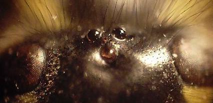
 Head of Polistes
A simple eye (sometimes called a pigment pit[1][2]) refers to a type of eye design or optical arrangement that contains a single lens which detect light. A "simple eye" is so-called in distinction from a multi-lensed "compound eye", and is not necessarily at all simple in the usual sense of the word. The eyes of humans and large animals, and camera lenses are classed as "simple" because in both cases a single lens collects and focuses light onto the retina or film. Many insects have compound eyes consisting of multiple lenses (up to tens of thousands), each focusing light onto a small number of retinula cells.
The structure of an animal's eye is determined by the environment in which it lives, and the behavioural tasks it must fulfill in order to survive. Arthropods differ widely in the habitats in which they live, as well as their visual requirements for finding food or conspecifics, and avoiding predators. Consequently, an enormous variety of eye designs are found in arthropods: nature has repeatedly developed novel solutions to overcome visual problems or limitations (for a review of arthropod visual systems see Warrant, 2006).[3]
Contents
Ocelli or eye spots
Jellyfish, sea stars, and flatworms bear the simplest eyes, pigment spot ocelli, which have pigment distributed randomly and which have no additional structures such as a cornea and lens. The apparent eye color in these animals is therefore red or black.[4]
Many snails and slugs (gastropod mollusks) also have ocelli, either at the tip of the tentacles or at the base of the tentacles. However some other gastropods, such as the Strombidae, have much more sophisticated eyes.
Simple eyes in arthropods
Spider eyes
 This jumping spider's main ocelli (center pair) are very acute. The outer pair are "secondary eyes" and there are other pairs of secondary eyes on the sides and top of its head.
Spiders, which do not have compound eyes, have several, usually four, pairs of simple eyes, with each pair adapted for a specific task or tasks. There are principal, absent in those with only three pairs, and secondary eyes. Only the principal eyes have a retina that can be moved. The secondary eyes have a reflector at the back of the eyes. The light-sensitive part of the receptor cells is next to this so they get direct and reflected light. In hunting or jumping spiders for example, a forward-facing pair possesses the best resolution (and even telescopic components) in order to see the (often small) prey at a large distance. Night-hunting spiders' eyes are very sensitive in low light levels with a large aperture, f/0.58.[5]
Dorsal ocelli
The term "ocelli" (singular ocellus) is derived from the Latin oculus (eye), and literally means "little eye". Two distinct ocellus types exist:[6] dorsal ocelli (or simply "ocelli"), found in most insects, and lateral ocelli (or stemmata), which are found in the larvae of some insect orders. They are structurally and functionally very different. Simple eyes of other animals, e.g. cnidarians may also be referred to as 'ocelli', but again the structure and anatomy of these eyes is quite distinct from those of the dorsal ocelli of insects.
Dorsal ocelli are light-sensitive organs found on the dorsal (top-most) surface or frontal surface of the head of many insects (e.g. Hymenoptera (bees, ants, wasps, sawflies), Diptera (flies), Odonata (dragonflies, damselflies) and Orthoptera (grasshoppers, locusts)). The ocelli co-exist with the compound eyes, thus most insects possess two anatomically separate and functionally different visual pathways.
The number, form, and function of the dorsal ocelli varies markedly throughout insect orders. They tend to be larger and more strongly expressed in flying insects (particularly bees, wasps, dragonflies and locusts), where they are typically found as a triplet. Two lateral ocelli are directed to the left and right of the head respectively, while a central (median) ocellus is directed frontally. In some terrestrial insects (e.g. some ants and cockroaches), only two lateral ocelli are present: the median ocellus is absent. Note that the unfortunately labelled "lateral ocelli" here refers to the sideways-facing position of the ocelli, which are of the dorsal type. They should not be confused with the lateral ocelli of some insect larvae (see stemmata).
A dorsal ocellus consists of a lens element (cornea) and a layer of photoreceptors (rod cells). As noted above, ocelli vary widely among insect orders. The ocellar lens may be strongly curved (e.g. bees, locusts, dragonflies) or flat (e.g. cockroaches). The photoreceptor layer may (e.g. locusts) or may not (e.g. blowflies, dragonflies) be separated from the lens by a clear zone (vitreous humour). The number of photoreceptors also varies widely, but may number in the hundreds or thousands for well developed ocelli.
Two somewhat unusual features of the ocelli are particularly notable and generally well conserved between insect orders.
- The refractive power of the lens is not typically sufficient to form an image on the photoreceptor layer.
- Dorsal ocelli ubiquitously have massive convergence ratios from first-order (photoreceptor) to second-order neurons.
These two factors have led to the conclusion that the dorsal ocelli are incapable of perceiving form, and are thus solely suitable for light metering functions. Given the large aperture and low f-number of the lens, as well as high convergence ratios and synaptic gains, the ocelli are generally considered to be far more sensitive to light than the compound eyes. Additionally, given the relatively simple neural arrangement of the eye (small number of synapses between detector and effector) as well as the extremely large diameter of some ocellar interneurons (often the largest diameter neurons in the animal's nervous system) the ocelli are typically considered to be "faster" than the compound eyes.[7]
One common theory of ocellar function in flying insects holds that they are used to assist in maintaining flight stability. Given their underfocused nature, wide fields of view, and high light collecting ability, the ocelli are superbly adapted for measuring changes in the perceived brightness of the external world as an insect rolls or pitches around its body axis during flight. Corrective flight responses to light have been demonstrated in locusts[8] and dragonflies[9] in tethered flight. Other theories of ocellar function have ranged from roles as light adaptors or global excitatory organs, polarization sensors, and circadian entrainers.
Recent studies have shown that the ocelli of some insects (most notably the dragonfly, but also some wasps) are capable of form vision as the ocellar lens forms an image within, or close to the photoreceptor layer.[3][10] In dragonflies it has been demonstrated that the receptive fields of both the photoreceptors[11] and the second-order neurons[12] can be quite restricted. Further research has demonstrated that these eyes not only resolve spatial details of the world, they also perceive motion.[13] Second-order neurons in the dragonfly median ocellus respond more strongly to upwards moving bars and gratings than to downwards moving bars and gratings. However this effect is only present when ultraviolet light is used in the stimulus; when ultraviolet light is absent, no directional response is observed. Dragonfly ocelli are especially highly developed and specialised visual organs, which may support the exceptional acrobatic abilities of these animals.
Research on the ocelli is of high interest to designers of small unmanned aerial vehicles. Designers of these craft face many of the same challenges that insects face in maintaining stability in a three-dimensional world. Engineers are increasingly taking inspiration from insects in order to overcome these challenges.[14]
Stemmata
Stemmata (singular stemma, also referred to as lateral ocelli) are the only eyes of the larvae of several orders of insects (fleas, springtails, silverfish, and Strepsiptera). They have a similar form to ommatidia, the constituent elements of compound eyes: thus they may not be considered as simple eyes. Behind each lens lies a single cluster of photoreceptor cells, termed a retinula. Their lens is biconvex, and their body contains a vitreous or crystalline core. They may represent simplified compound eyes, reflected by their lateral position on the head. They are possessed by myriapods and some insect larvae.[6]
Genetic controls
A number of genetic pathways are responsible for the occurrence and positioning of the ocelli. The gene orthodenticle is allelic to ocelliless, a mutation that stops ocelli being produced.[15] In Drosophila, the rhodopsin Rh2 is only expressed in simple eyes.[16]
Whilst (in Drosophila at least) the genes eyeless and daschund are both expressed in the compound eye but not the simple eye, there are no reported developmental genes uniquely expressed in the simple eye.[17]
Egfr (Epidermal growth factor receptor) promotes the expression of orthodenticle (and possibly Eyes absent ('Eya')) and as such is essential for simple eye formation.[17]
See also
References
- ^ http://www.mendeley.com/research/structure-and-optics-of-the-eyes-of-the-box-jellyfish-chiropsella-bronzie/
- ^ . PMC 2825319. http://www.pubmedcentral.nih.gov/articlerender.fcgi?tool=pmcentrez&artid=2825319.
- ^ a b Eric J. Warrant, Almut Kelber, Rita Wallén & William T. Wcislo (December 2006). "Ocellar optics in nocturnal and diurnal bees and wasps". Arthropod Structure & Development 35 (4): 293–305. doi:10.1016/j.asd.2006.08.012. PMID 18089077.
- ^ "Eye (invertebrate)". McGraw-Hill Encyclopedia of Science & Technology. 6. 2007. p. 790.
- ^ Blest, AD; Land (1997). "The Physiological optics of Dinopis Subrufus L.Koch: a fisheye lens in a spider". Proceedings of the Royal Society, London (196): 198–222.
- ^ a b C. Bitsch & J. Bitsch (2005). "Evolution of eye structure and arthropod phylogeny". In Stefan Koenemann & Ronald Jenner. Crustacea and Arthropod Relationships. Volume 16 of Crustacean Issues. Taylor & Francis. pp. 185–214. ISBN 9780849334986. http://books.google.com/?id=7ZHtG3aELesC&printsec=frontcover&dq=evolution+of+the+eye+review#PPA185,M1.
- ^ Martin Wilson (1978). "The functional organisation of locust ocelli". Journal of Comparative Physiology A: Neuroethology, Sensory, Neural, and Behavioral Physiology 124 (4): 297–316. doi:10.1007/BF00661380. http://www.springerlink.com/content/x0j046w843352w63/.
- ^ Charles P. Taylor (1981). "Contribution of compound eyes and ocelli to steering of locusts in flight: I. Behavioural analysis". Journal of Experimental Biology 93 (1): 1–18. http://jeb.biologists.org/cgi/content/abstract/93/1/1.
- ^ Gert Stange & Jonathon Howard (1979). "An ocellar dorsal light response in a dragonfly". Journal of Experimental Biology 83 (1): 351–355. http://jeb.biologists.org/cgi/reprint/83/1/351.
- ^ Richard P. Berry, Gert Stange & Eric J. Warrant (May 2007). "Form vision in the insect dorsal ocelli: an anatomical and optical analysis of the dragonfly median ocellus". Vision Research 47 (10): 1394–1409. doi:10.1016/j.visres.2007.01.019. PMID 17368709.
- ^ Joshua van Kleef, Andrew Charles James & Gert Stange (October 2005). "A spatiotemporal white noise analysis of photoreceptor responses to UV and green light in the dragonfly median ocellus". Journal of General Physiology 126 (5): 481–497. doi:10.1085/jgp.200509319. PMC 2266605. PMID 16260838. http://www.pubmedcentral.nih.gov/articlerender.fcgi?tool=pmcentrez&artid=2266605.
- ^ Richard Berry, Joshua van Kleef & Gert Stange (May 2007). "The mapping of visual space by dragonfly lateral ocelli". Journal of Comparative Physiology A: Neuroethology, Sensory, Neural, and Behavioral Physiology 193 (5): 495–513. doi:10.1007/s00359-006-0204-8. PMID 17273849.
- ^ Joshua van Kleef, Richard Berry & Gert Stange (March 2008). "Directional selectivity in the simple eye of an insect". The Journal of Neuroscience 28 (11): 2845–2855. doi:10.1523/JNEUROSCI.5556-07.2008. PMID 18337415.
- ^ Gert Stange, R. Berry & J. van Kleef (September 2007). "Design concepts for a novel attitude sensor for Micro Air Vehicles, based on dragonfly ocellar vision". 3rd US-European Competition and Workshop on Micro Air Vehicle Systems (MAV07) & European Micro Air Vehicle Conference and Flight Competition (EMAV2007) 1: 17–21.
- ^ R. Finkelstein, D. Smouse, T. M. Capaci, A. C. Spradling & N Perrimon (1990). "The orthodenticle gene encodes a novel homeo domain protein involved in the development of the Drosophila nervous system and ocellar visual structures". Genes & Development 4: 1516–1527. doi:10.1101/gad.4.9.1516.
- ^ Adriana D. Briscoe & Lars Chittka (2001). "The evolution of color vision in insects". Annual Review of Entomology 46: 471–510. doi:10.1146/annurev.ento.46.1.471. PMID 11112177.
- ^ a b Markus Friedrich (2006). "Ancient mechanisms of visual sense organ development based on comparison of the gene networks controlling larval eye, ocellus, and compound eye specification in Drosophila". Arthropod Structure & Development 35 (4): 357–378. doi:10.1016/j.asd.2006.08.010. PMID 18089081.
Further reading
Warrant, Eric; Dan-Eric Nilsson (2006). Invertebrate Vision. Cambridge University Press. ISBN 978-0521830881. http://www.cambridge.org/catalogue/catalogue.asp?isbn=9780521830881&ss=fro.
External links
- John R. Meyer, Photoreceptors
Eyes - Arthropod eye
- Cephalopod eye
- Eye shine
- Gastropod eye
- Human eye
- Simple eye in invertebrates
- Mammalian eye
- Mollusc eye
- Parietal eye
Evolution Colouration - Animal colouration
- Aposematism
- Camouflage
- Chromatophore
- Countershading
- Crypsis
- Eyespot (mimicry)
- Theory of camouflage
- Underwater camouflage and mimicry
Related topics Categories:- Eye
- Arthropod anatomy


Wikimedia Foundation. 2010.