- Optic vesicle
-
Optic vesicle 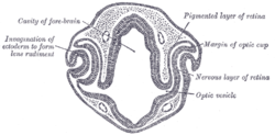
Transverse section of head of chick embryo of forty-eight hours’ incubation. (Optic vesicle labeled at lower right.) 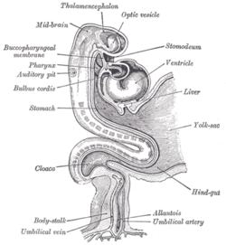
Human embryo about fifteen days old. Brain and heart represented from right side. Digestive tube and yolk sac in median section. (Optic vesicle labeled at center top.) Latin vesicula optica; vesicula ophthalmica Gray's subject #224 1001 Carnegie stage 11 Code TE E5.14.3.4.2.2.4 The eyes begin to develop as a pair of diverticula from the lateral aspects of the forebrain. These diverticula make their appearance before the closure of the anterior end of the neural tube; after the closure of the tube they are known as the optic vesicles.
They project toward the sides of the head, and the peripheral part of each expands to form a hollow bulb, while the proximal part remains narrow and constitutes the optic stalk.
Additional images
-

Head of chick embryo of about thirty-eight hours’ incubation, viewed from the ventral surface. X 26
External links
This article was originally based on an entry from a public domain edition of Gray's Anatomy. As such, some of the information contained within it may be outdated.
Prenatal development/Mammalian development of nervous system (GA 9.733 and GA 10.1002, TE E5.13-16) Neurogenesis Cranial neural crest (Cardiac neural crest complex) · Truncal neural crestRostral neuropore
Cephalic flexure · Pontine flexure
Alar plate (sensory) · Basal plate (motor)
Germinal matrixEye development Auditory development M: EYE
anat(g/a/p)/phys/devp/prot
noco/cong/tumr, epon
proc, drug(S1A/1E/1F/1L)
M: EAR
anat(e/p)/phys/devp
noco/cong, epon
proc, drug(S2)
Categories:- Eye stubs
- Embryology of nervous system
- Eye
-


Wikimedia Foundation. 2010.