- Cardiac cycle
-
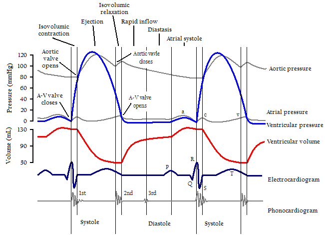
 Cardiac events occurring in the cardiac cycle. Two complete cycles are illustrated.
The cardiac cycle is a term referring to all or any of the events related to the flow or blood pressure that occurs from the beginning of one heartbeat to the beginning of the next.[1] The frequency of the cardiac cycle is described by the heart rate. Each beat of the heart involves five major stages. The first two stages, often considered together as the "ventricular filling" stage, involve the movement of blood from atria into ventricles. The next three stages involve the movement of blood from the ventricles to the pulmonary artery (in the case of the right ventricle) and the aorta (in the case of the left ventricle).[1]
The first, "early diastole", is when the semilunar valves close, the atrioventricular (AV) valves open, and the whole heart is relaxed. The second, "atrial systole", is when the atrium contracts, the AV valves open, and blood flows from atrium to the ventricle. The third, "isovolumic ventricular contraction", is when the ventricles begin to contract, the AV and semilunar valves close, and there is no change in volume. The fourth, "ventricular ejection", is when the ventricles are empty and contracting, and the semilunar valves are open. During the fifth stage, "Isovolumic ventricular relaxation", pressure decreases, no blood enters the ventricles, the ventricles stop contracting and begin to relax, and the semilunar valves close due to the pressure of blood in the aorta.
Throughout the cardiac cycle, blood pressure increases and decreases. The cardiac cycle is coordinated by a series of electrical impulses that are produced by specialized heart cells found within the sinoatrial node and the atrioventricular node. The cardiac muscle is composed of myocytes which initiate their own contraction without help of external nerves (with the exception of modifying the heart rate due to metabolic demand). Under normal circumstances, each cycle takes approximately one second.
Contents
Anatomical basis of the cardiac cycle
Main article: HeartThe heart is a four-chambered organ consisting of right and left halves. Two of the chambers, the left and right atria, are entry-points into the heart, while the other two chambers, the left and right ventricles, are responsible for contractions that send the blood through the circulation. The circulation is split into the pulmonary and systemic circulation. The right ventricle's role is to pump deoxygenated blood to the lungs through the pulmonary trunk and pulmonary arteries. The left ventricle's role is to pump now oxygenated blood to the body through the aorta.
Importantly, the right and left ventricles contract simultaneously, and so in consideration of the cardiac cycle the events that are occurring on one side of the heart are equivalent to the events occurring on the other side of the heart. However, the ventricles contract shortly after the atria. The sino-atrial node sends out electrical waves of excitation to both atria, and it is prevented from flowing into the ventricles by strands of non-conducting fibrous tissue situated laterally from the tricuspid/bicuspid valves to the septum. These waves of excitation travel towards the septum and into the atrio-ventricular node, where they are held for roughly 0.1 seconds. They are then discharged down the bundle of his, then down the purkinje tissue, which are both situated inside the septum. The waves flow down towards the apex of the heart and are then discharged into the ventricles, causing them to contract (ventricular systole) This creates the well known beat of the heart.
Atrial systole
 Atrial systole
Atrial systole is the contraction of the heart muscle (myocardia) of the left and right atria. Normally, both atria contract at the same time. The term systole is synonymous with contraction (movement or shortening) of a muscle. Electrical systole is the electrical activity that stimulates the myocardium of the chambers of the heart to make them contract. This is soon followed by Mechanical systole, which is the mechanical contraction of the heart.
As the atria contract, the blood pressure in each atrium increases, forcing additional blood into the ventricles. The additional flow of blood is called atrial kick.
80% of the blood flows passively down to the ventricles, so the atria do not have to contract a great amount.[2]
Atrial kick is absent if there is loss of normal electrical conduction in the heart, such as during atrial fibrillation, atrial flutter, and complete heart block. Atrial kick is also different in character depending on the condition of the heart, such as stiff heart, which is found in patients with diastolic dysfunction.
Detection of atrial systole
Electrical systole of the atria begins with the onset of the P wave on the ECG. The wave of bipolarization (or depolarization) that stimulates both atria to contract at the same time is due to sinoatrial node which is located on the upper wall of the right atrium.
Ventricular systole
 Ventricular systole
Ventricular systole is the contraction of the muscles (myocardia) of the left and right ventricles.
At the later part of the ejection phase, although the ventricular pressure falls below the aortic pressure, the aortic valve remains patent because of the inertial energy of the ejected blood.[3]
The graph of aortic pressure throughout the cardiac cycle displays a small dip (the "incisure" or "dicrotic notch") which coincides with the aortic valve closure. The dip in the graph is immediately followed by a brief rise (the "dicrotic wave") then gradual decline. Just as the ventricles enter into diastole, the brief reversal of flow from the aorta back into the left ventricle causes the aortic valves to shut. This results in the slight increase in aortic pressure caused by the elastic recoil of the semilunar valves and aorta.[4][5][6]
Detection of ventricular systole
Heart sounds
Main article: Heart soundsThe closing of the mitral and tricuspid valves (known together as the atrioventricular valves) at the beginning of ventricular systole cause the first part of the "lubb-dubb" sound made by the heart as it beats. Formally, this sound is known as the First Heart Tone, or S1. This first heart tone is created by the closure of mitral and tricuspid valve and is actually a two component sound, M1, T1.
The second part of the "lub-dubb" (the Second Heart Tone, or S2), is caused by the closure of the aortic and pulmonary valves at the end of ventricular systole. As the left ventricle empties, its pressure falls below the pressure in the aorta, and the aortic valve closes. Similarly, as the pressure in the right ventricle falls below the pressure in the pulmonary artery, the pulmonary valve closes. The second heart sound is also two components, A2 and P2. The aortic valve closes earlier than the pulmonary valve and they are audibly separated from each other in the second heart sound. This "splitting" of S2 is only audible during inhalation. However, some cardiac conduction abnormalities such as left bundle branch block (LBBB) allow the P2 sound to be heard before the A2 sound during expiration. With LBBB, inhalation brings A2 and P2 closer together where they cannot be audibly distinguished.
Electrocardiogram
In an electrocardiogram, electrical systole of the ventricles begins at the beginning of the QRS complex.
Diastole
Cardiac diastoleCardiac Diastole is the period of time when the heart relaxes after contraction in preparation for refilling with circulating blood. Ventricular diastole is when the ventricles are relaxing, while atrial diastole is when the atria are relaxing. Together they are known as complete cardiac diastole.
During ventricular diastole, the pressure in the (left and right) ventricles drops from the peak that it reaches in systole. When the pressure in the left ventricle drops to below the pressure in the left atrium, the mitral valve opens, and the left ventricle fills with blood that was accumulating in the left atrium. The isovolumic relaxation time (IVRT) is the interval from the aortic component of the second heart sound, that is, closure of the aortic valve, to onset of filling by opening of the mitral valve.[7] Likewise, when the pressure in the right ventricle drops below that in the right atrium, the tricuspid valve opens, and the right ventricle fills with blood that was accumulating in the right atrium. During diastole the pressure within the right ventricle is lower than that in aorta, allowing blood to circulate in the heart itself via the coronary arteries.
Regulation of the cardiac cycle
Cardiac muscle has automaticity, which means that it is self-exciting. (You could also call it "myogenic" tissue. Meaning a tissue able of creating its own excitement.) This is in contrast with skeletal muscle, which requires either conscious or reflex nervous stimuli for excitation. The heart's rhythmic contractions occur spontaneously, although the rate of contraction can be changed by nervous or hormonal influences, exercise and emotions. For example, the sympathetic nerves to accelerate heart rate and the vagus nerve decelerates heart rate.
The rhythmic sequence of contractions is coordinated by the sinoatrial (SA) and atrioventricular (AV) nodes. The sinoatrial node, often known as the cardiac pacemaker, is located in the upper wall of the right atrium and is responsible for the wave of electrical stimulation that initiates atrial contraction by creating an action potential. Once the wave reaches the AV node, situated in the lower right atrium, it is delayed there before being conducted through the bundles of His and back up the Purkinje fibers, leading to a contraction of the ventricles. The delay at the AV node allows enough time for all of the blood in the atria to fill their respective ventricles. In the event of severe pathology, the AV node can also act as a pacemaker; this is usually not the case because their rate of spontaneous firing is considerably lower than that of the pacemaker cells in the SA node and hence is overridden.
See also
- Apex beat
- Blood pressure
- Cardiac action potential
- Cardiac muscle
- Cardiac output
- Electrocardiogram
- Heart
- Systolic array (computer architecture)
- Ventricle
References
- ^ a b Guyton, A.C. & Hall, J.E. (2006) Textbook of Medical Physiology (11th ed.) Philadelphia: Elsevier Saunder ISBN 0-7216-0240-1
- ^ Advanced Biology for You - Gareth Williams
- ^ Cardiovascular Physiology Concepts by Richard E. Klabunde, Ph.D.: Cardiac Cycle - Reduced Ejection (Phase 4)
- ^ Plethysmograph
- ^ The Heart
- ^ Human Cardiopulmonary Physiology
- ^ Inductance cardiography (thoracocardiography): A novel, noninvasive technique for monitoring left ventricular filling. Journal of Critical Care, Volume 14, Issue 4, Pages 177-185
External links
- Interactive cardiac cycle. Interactivephysiology.com
- Cardiac cycle. University of Utah School of Medicine
- Heart sounds. UCLA School of Medicine
Categories:

Wikimedia Foundation. 2010.