- Craniofacial surgery
-
Craniofacial surgery is a surgical subspecialty of maxillofacial surgery, plastic surgery, and ENT that deals with congenital and acquired deformities of the skull, face, and jaws. Although craniofacial treatment often involves manipulation of bone, craniofacial surgery is not tissue-specific, i.e., craniofacial surgeons deal with bone, skin, muscle, teeth, etc. Craniofacial surgery does not, however, include surgery of the brain or eye.
Defects typically treated by craniofacial surgeons include craniosynostosis (isolated and syndromic), rare craniofacial clefts, acute and chronic sequellae of facial fractures, cleft lip and palate, micrognathia, Treacher Collins Syndrome, Apert's Syndrome, Crouzon's Syndrome, hemifacial microsomia and many others.
Training in craniofacial surgery usually consists of a 1-year surgical fellowship completed after a residency in either plastic surgery, oral and maxillofacial surgery, or otolaryngology.
Contents
Craniosynostosis
Main article: Craniosynostosis Fig. 1 Cranial sutures viewed from top of head
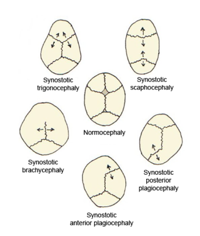
 Fig. 2 Skull deformities associated with single suture synostosis
The bones of the human skull are joined together by cranial sutures (see figure 1). The anterior fontanelle is where the metopic, saggital and coronal sutures meet. Normally the sutures gradually fuse within the first few years after birth. In infants where one or more of the sutures fuses too early the growth of the skull is restricted, resulting in compensation mechanisms which cause irregular growth patterns. Growth in the skull is perpendicular to the sutures. When a suture fuses too early, the growth perpendicular to that suture will be restricted, and the bone growth near the other sutures will be stimulated, causing an abnormal head shape. The expanding brain is the main stimulus for the rapid growth of the skull in the first years of life. Inhibited growth potential of the skull can restrict the volume, needed by the brain. In cases in which the compensation does not effectively provide enough space for the growing brain, craniosynostosis results in increased intracranial pressure.[1]
Craniosynostosis is called simple when one suture is involved, and complex when two or more sutres are involved. It can occur as part of a syndrome or as an isolated defect (nonsyndromic).[2]
There are several classifications of deformities of the human skull, we will discuss them in order of prevalence.
Scaphocephaly
In scaphocephaly the saggital suture is prematurely fused. The saggital suture runs from the front to the back of the head. The shape of this deformity is a long narrow head, formed like a boat (greek skaphe, “light boat or skiff”). The incidence of scaphocephaly is 2.8 per 10 000 births in the Netherlands and is therefore the most common form of craniosynostosis.[3][4]
Trigonocephaly
In trigonocephaly the metopic suture is prematurely fused. The metopic suture is situated in the medial line of the forehead. Premature fusion of this suture caused the forehead to become pointed, giving the head a triangular shape when viewd from above (greek trigono, “triangle”). The incidence of trigonocephaly is 1 - 1.9 per 10 000 births in the Netherlands.[3]
Plagiocephaly
In plagiocephaly one of the coronal sutures is prematurely fused. The coronal sutures run over the top of the head, just in front of the ears. The shape of this deformity is an asymmetrical distortion (flattening of one side of the head) as you can see in figure 2. The incidence is 1 in 10 000 births. [3][5]
Brachycephaly
In brachycephaly both of the coronal sutures are prematurely fused. The shape of this deformity is a wide and high head. The incidence at birth is 1/20 000.[6]
Surgical procedures
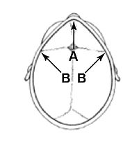 Fig. 3 Locations of the incisions used in fronto-supraorbital advancement.
In cases where the forehead is involved (trigonocephaly and plagiocephaly), a technique called fronto-supraorbital advancement is used to correct the shape of the head. The procedure is performed at a young age in order to provide the brain with enough space to grow and prevent further abnormal growth of the skull. Fronto-orbital advancement literally means moving the front of the skull including the eye sockets forward. A section of the skull, ranging from the coronal sutures to the eye sockets is cut loose in order to correct the shape of the skull. The incision is cut in a zigzag shape from ear to ear so that the hair will cover the scar and make it less visible. The incision is made to the bone only, leaving the underlying meninges intact. The top half of the eye sockets is cut loose. Once the eye socket section has been cut loose, a vertical incision is made in the midline, and the whole section of the eye socket is bent outwards in order to correct the pointed shape of the forehead. Because the section is now too wide, a wedge needs to be cut on either side to allow the section to fit into the skull. Figure 4 shows the sections that are loosened and adjusted, and figure 3 shows the location of the vertical incision (arrow A) and the two wedges (arrow B).
In scaphocephaly the saggital suture is prematurely fused, preventing the skull from growing perpendicular to the suture. Thus the head becomes very narrow and long. If a scaphocephaly is diagnosed within 4 to 5 months after birth, it can be corrected with a relatively simple procedure whereby the saggital suture is surgically reopened. Once the suture has been opened the bone segments will be able to grow again and the head can regain its normal shape. This operation is only performed on patients younger than five months old with a scaphocephaly. This is due to the fact that the bone segments only have the ability to adapt so severely when the operation is performed at this young age. A scaphocephaly that is diagnosed and treated later in life requires a more extensive secondary operation than one which is treated before five months.
 Fig. 4 Bone segments that are removed in fronto-supraorbital advancement
Ethical considerations
The Hastings Center, a prominent bioethics research institute, conducted a project called "Surgically Shaping Children". The project produced an edited volume (Parens, 2006) which considers reconstructive surgery on children with craniofacial anomalies, ambiguous genitalia, and achondroplasia.
See also
- Oral and maxillofacial surgery
- Plastic surgery
- Scalp reconstruction
External links
- Information about Craniofacial Surgery for Children and Adults
- BAOMS - What is craniofacial surgery?
- [1]
- Journal of Craniofacial Surgery
- American Society of Craniofacial Surgery
- European Association of Cranio-Maxillofacial Surgery
- International Society of Craniofacial Surgery
- Cleft and Craniofacial Center at Hasbro Children's Hospital
Further reading
Parens, E., Ed. (2006). Surgically Shaping Children : Technology, Ethics, and the Pursuit of Normality. Baltimore, Johns Hopkins University Press. ISBN 0-8018-8305-9.
References
- ^ JJ van der Vlugt, JJ van der Meulen and HE Creemers, et al., The risk of psychopathology in children with craniosynostosis, Plastic and Reconstructive Surgery, December 2009, pp 2054-2060
- ^ www.aafp.org/afp/2004/0615/p2863.html
- ^ a b c CF Kweldam, JJ van der Vlugt and JJNM van der Meulen, The incidence of craniosynostosis in the Netherlands 1997 – 2007, Journal of Plastic, Reconstructive & Aesthetic Surgery
- ^ BL Hutchison, Alistair W Stewart and Edwin A Mitchell, Characteristics, head shape measurements and developmental delay in 287 consecutive infants attending a plagiocephaly clinic, Acta Pædiatrica98, September 2009, pp 1494–1499
- ^ www.orpha.net
- ^ www.orpha.net
Categories:


Wikimedia Foundation. 2010.