- Trapezius muscle
-
This article is about the human skeletal muscle. For the trapezius muscles found in cats, see trapezius muscles (cat).
Trapezius 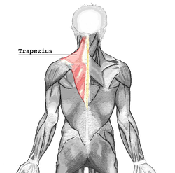
trapezius 
muscles connecting the upper extremity to the vertebral column; trapezius is labeled at upper left. Latin musculus trapezius Gray's subject #121 432 Origin external occipital protuberance, nuchal ligament, medial superior nuchal line, spinous processes of vertebrae C7-T12 Insertion posterior border of the lateral third of the clavicle, acromion process, and spine of scapula Artery transverse cervical artery [1] Nerve accessory nerve (motor)
cervical spinal nerves C3 and C4 (motor and sensation)Actions rotation, retraction, elevation, and depression of scapula Antagonist serratus anterior muscle, Latissimus dorsi In human anatomy, the trapezius is a large superficial muscle that extends longitudinally from the occipital bone to the lower thoracic vertebrae and laterally to the spine of the scapula (shoulder blade). Its functions are to move the scapulae and support the arm.
The trapezius has three functional regions: the superior region (descending part), which supports the weight of the arm; the intermediate region (transverse part), which retracts the scapulae; and the inferior region (ascending part), which medially rotates and depresses the scapulae.
Contents
Terminology
The trapezius muscle resembles a trapezium (trapezoid in American English), or diamond-shaped quadrilateral. The word "spinotrapezius" refers to the human trapezius, although it is not commonly used in modern texts. In other mammals, it refers to a portion of the analogous muscle.
Anatomy
The superior or upper fibers of the trapezius arise from the external occipital protuberance, the medial third of the superior nuchal line of the occipital bone (both in the back of the head), and the ligamentum nuchae. From this origin they proceed downward and laterally to be inserted into the posterior border of the lateral third of the clavicle.
The middle fibers of the trapezius arise from the spinous process of the seventh cervical (both in the back of the neck), and the spinous processes of the first, second and third thoracic vertebrae. They are inserted into the medial margin of the acromion, and into the superior lip of the posterior border of the spine of the scapula.
The inferior or lower fibers of the trapezius arise from the spinous processes of the remaining thoracic vertebrae (T4-T12). From this origin they proceed upward and laterally to converge near the scapula and end in an aponeurosis, which glides over the smooth triangular surface on the medial end of the spine, to be inserted into a tubercle at the apex of this smooth triangular surface.
At its occipital origin, the trapezius is connected to the bone by a thin fibrous lamina, firmly adherent to the skin. The superficial and deep epimysia are continuous with an investing deep fascia that encircles the neck and also contains both sternocleidomastoid muscles.
At the middle, the muscle is connected to the spinous processes by a broad semi-elliptical aponeurosis, which reaches from the sixth cervical to the third thoracic vertebræ and forms, with that of the opposite muscle, a tendinous ellipse. The rest of the muscle arises by numerous short tendinous fibers.
Innervation
Motor function is supplied by the accessory nerve (CN XI). Sensation, including pain and proprioception, travel via the ventral rami of the third (C3) and fourth (C4) cervical nerves. Since it is a muscle of the upper limb, the trapezius is not innervated by dorsal rami despite being placed superficially in the back.
It is possible to feel the muscles of the superior trapezius become active by holding a weight in one hand in front of the body and, with the other hand, touching the area between the shoulder and the neck.
Actions and exercises
Contraction of the trapezius muscle can have two effects: movement of the scapulae when the spinal origins are stable, and movement of the spine when the scapulae are stable.
Scapular movements
The upper portion of the trapezius can be developed by elevating the shoulders. Common exercises for this movement are shoulder shrugs and upright rows. Middle fibers are developed by pulling shoulder blades together. The lower part can be developed by drawing the shoulder blades downward while keeping the arms almost straight and stiff. It is mainly used in throwing, with the deltoid muscles.
The upper and lower trapezius fibers also work in tandem with the serratus anterior to upwardly rotate the scapulae, such as during an overhead press. When activating together, the upper and lower fibers also assist the middle fibers (along with other muscles such as the rhomboids) with scapular retraction/adduction.
Spinal movements
When the scapulae are stable (such as lying on one's back, or using muscles to fix them in space), trapezius contractions can cause spinal movements, particularly the upper fibers on the cervical vertebrae, a co-contraction of both sides can extend the neck.
Balance
Muscle imbalances, which can heavily affect posture and compromise shoulder health, can result if all three sections of the trapezius are not developed equally.[2]
References
- ^ "Tufts". http://iris3.med.tufts.edu/headneck/Triangles/Posterior%20Triangle%20of%20the%20Neck.htm. Retrieved 2007-12-11.
- ^ Griffin, John C. (2006). Client-Centered Exercise Prescription. Champaign, IL: Human Kinetics. p. 217. ISBN 978-0-7360-5495-9. http://books.google.com/books?id=qkQNO_3vb6oC.
Dissection Video
External links
This article was originally based on an entry from a public domain edition of Gray's Anatomy. As such, some of the information contained within it may be outdated.
Back splenius (capitis, cervicis) · erector spinae (iliocostalis, longissimus, spinalis) · latissimus dorsi
transversospinales: (semispinalis dorsi, semispinalis cervicis, semispinalis capitis, multifidus, rotatores) · interspinales · intertransversarii
Vertebral column: trapezius · latissimus dorsi · rhomboid (major, minor) · levator scapulae
fascia: Thoracolumbar fasciaThorax intercostales (external, internal, innermost) · subcostales · transversus thoracis · levatores costarum · serratus posterior (inferior, superior) · diaphragm
Thoracic cavity: pectoralis major · pectoralis minor · subclavius · serratus anterior
fascia: Pectoral fascia · Clavipectoral fasciaCategories:- Back anatomy
- Muscles of the upper limb


Wikimedia Foundation. 2010.