- Mast cell
-
Mast cell 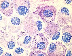
Mast cells Latin mastocytus Code TH H2.00.03.0.01010 A mast cell (also known as mastocyte and labrocyte[1]) is a resident cell of several types of tissues and contains many granules rich in histamine and heparin. Although best known for their role in allergy and anaphylaxis, mast cells play an important protective role as well, being intimately involved in wound healing and defense against pathogens.[2]
Contents
Origin and classification
 Mast cell
Mast cells were first described by Paul Ehrlich in his 1878 doctoral thesis on the basis of their unique staining characteristics and large granules. These granules also led him to the mistaken belief that they existed to nourish the surrounding tissue, and he named them "Mastzellen" (from the German: Mast, "food").[3][4] They are now considered to be part of the immune system.
Mast cells are very close to basophil granulocytes (a class of white blood cells) in blood; the similarities between mast cells and basophils have led many to speculate that mast cells are basophils that have "homed in" on tissues. However, current evidence suggests that they are generated by different precursor cells in the bone marrow. Nevertheless, both mast cells and basophils are thought to originate from bone marrow precursors expressing the CD34 molecule. Basophils leave the bone marrow already mature, whereas the mast cell circulates in an immature form, only maturing once in a tissue site. The site an immature mast cell settles in probably determines its precise characteristics.[2]
Two types of mast cells are recognized, those from connective tissue and a distinct set of mucosal mast cells. The activities of the latter are dependent on T-cells.[5]
Mast cells are present in most tissues characteristically surrounding blood vessels and nerves, and are especially prominent near the boundaries between the outside world and the internal milieu, such as the skin, mucosa of the lungs and digestive tract, as well as in the mouth, conjunctiva and nose.[2]
Physiology
Mast cells play a key role in the inflammatory process. When activated, a mast cell rapidly releases its characteristic granules and various hormonal mediators into the interstitium. Mast cells can be stimulated to degranulate by direct injury (e.g. physical or chemical [such as opioids, alcohols, and certain antibiotics such as polymyxins]), cross-linking of Immunoglobulin E (IgE) receptors, or by activated complement proteins.[2]
Mast cells express a high-affinity receptor (FcεRI) for the Fc region of IgE, the least-abundant member of the antibodies. This receptor is of such high affinity that binding of IgE molecules is essentially irreversible. As a result, mast cells are coated with IgE. IgE is produced by Plasma cells (the antibody-producing cells of the immune system). IgE molecules, like all antibodies, are specific to one particular antigen.
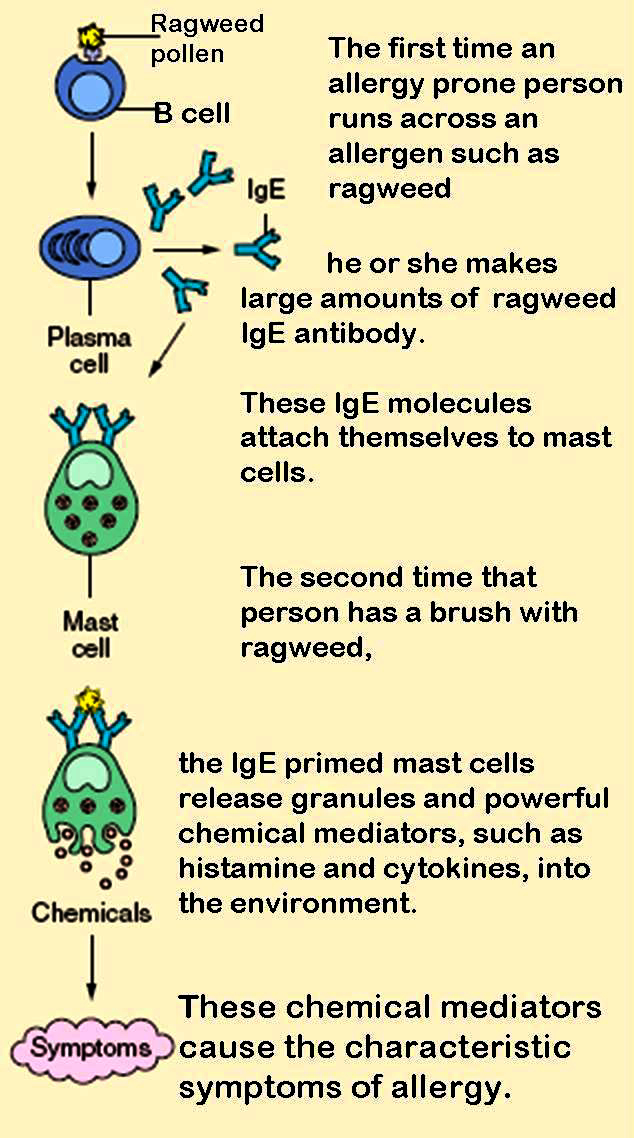
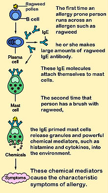 The role of mast cells in the development of allergy.
In allergic reactions, mast cells remain inactive until an allergen binds to IgE already in association with the cell (see above). Other membrane activation events can either prime mast cells for subsequent degranulation or can act in synergy with FceRI signal transduction.[6] Allergens are generally proteins or polysaccharides. The allergen binds to the antigen-binding sites, which are situated on the variable regions of the IgE molecules bound to the mast cell surface. It appears that binding of two or more IgE molecules (cross-linking) is required to activate the mast cell. The clustering of the intracellular domains of the cell-bound Fc receptors, which are associated with the cross-linked IgE molecules, causes a complex sequence of reactions inside the mast cell that lead to its activation. Although this reaction is most well understood in terms of allergy, it appears to have evolved as a defense system against intestinal worm infestations (tapeworms, etc.)[citation needed].
The molecules thus released into the extracellular environment include:[2]
- preformed mediators (from the granules):
- serine proteases, such as tryptase
- histamine (2-5 pg/cell)
- serotonin
- proteoglycans, mainly heparin (active as anticoagulant)
- newly formed lipid mediators (eicosanoids):
- cytokines
- Eosinophil chemotactic factor
Histamine dilates post capillary venules, activates the endothelium, and increases blood vessel permeability. This leads to local edema (swelling), warmth, redness, and the attraction of other inflammatory cells to the site of release. It also irritates nerve endings (leading to itching or pain). Cutaneous signs of histamine release are the "flare and wheal"-reaction. The bump and redness immediately following a mosquito bite are a good example of this reaction, which occurs seconds after challenge of the mast cell by an allergen.[2]
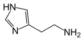 Structure of histamine
The other physiologic activities of mast cells are much less well-understood. Several lines of evidence suggest that mast cells may have a fairly fundamental role in innate immunity – they are capable of elaborating a vast array of important cytokines and other inflammatory mediators such as TNFa, they express multiple "pattern recognition receptors" thought to be involved in recognizing broad classes of pathogens, and mice without mast cells seem to be much more susceptible to a variety of infections.[citation needed]
Mast cell granules carry a variety of bioactive chemicals. These granules have been found to be transferred to adjacent cells of the immune system and neurons via transgranulation via their pseudopodia.[7]
Role in disease
Allergic disease
Many forms of cutaneous and mucosal allergy are mediated for a large part by mast cells; they play a central role in asthma, eczema, itch (from various causes) and allergic rhinitis and allergic conjunctivitis. Antihistamine drugs act by blocking the action of histamine on nerve endings. Cromoglicate-based drugs (sodium cromoglicate, nedocromil) block a calcium channel essential for mast cell degranulation, stabilizing the cell and preventing release of histamine and related mediators. Leukotriene antagonists (such as montelukast and zafirlukast) block the action of leukotriene mediators, and are being used increasingly in allergic diseases.[2]
Anaphylaxis
In anaphylaxis (a severe systemic reaction to allergens, such as nuts, bee stings or drugs), body-wide degranulation of mast cells leads to vasodilation and, if severe, symptoms of life-threatening shock.[citation needed]
Autoimmunity
Mast cells are implicated in the pathology associated with the autoimmune disorders rheumatoid arthritis, bullous pemphigoid, and multiple sclerosis. They have been shown to be involved in the recruitment of inflammatory cells to the joints (e.g. rheumatoid arthritis) and skin (e.g. bullous pemphigoid) and this activity is dependent on antibodies and complement components.[8]
Reproductive disorders
Mast cells are present within the endometrium, with increased activation and release of mediators in endometriosis.[9] In males, mast cells are present in the testes and are increased in oligo- and azoospermia, with mast cell mediators directly suppressing sperm motility in a potentially reversible manner.[9]
Mast cell disorders
Mastocytosis is a rare condition featuring proliferation of mast cells. It exists in a cutaneous and systemic form, with the former being limited to the skin and the latter involving multiple organs.[2] Mast cell tumors are often seen in dogs and cats.[10]
Histological staining
Toluidine blue - one of the most common stains for acid mucopolysaccharides and glycoaminoglycans, components of mast cells granules.[11]
References
- ^ http://www.memidex.com/labrocytes
- ^ a b c d e f g h Prussin C, Metcalfe DD (2003). "IgE, mast cells, basophils, and eosinophils". J Allergy Clin Immunol 111 (2 Suppl): S486–94. doi:10.1067/mai.2003.120. PMID 12592295.
- ^ Ehrlich P. Beiträge zur Theorie und Praxis der histologischen Färbung. Dissertation at Leipzig University, 1878.
- ^ [1]
- ^ Denburg, Judah A. (1998). Allergy and allergic diseases: the new mechanisms and therapeutics. Totowa, NJ: Humana Press. ISBN 0-89603-404-6.
- ^ Pulendran B, Ono SJ (May 2008). "A shot in the arm for mast cells". Nat. Med. 14 (5): 489–90. doi:10.1038/nm0508-489. PMID 18463655.
- ^ Wilhelm M, Silver R, Silverman AJ (November 2005). "Central nervous system neurons acquire mast cell products via transgranulation". Eur. J. Neurosci. 22 (9): 2238–48. doi:10.1111/j.1460-9568.2005.04429.x. PMID 16262662.
- ^ http://www.sciencemag.org/cgi/content/full/297/5587/1689
- ^ a b Menzies, F. M.; Shepherd, M. C.; Nibbs, R. J.; Nelson, S. M. (2010). "The role of mast cells and their mediators in reproduction, pregnancy and labour". Human Reproduction Update 17 (3): 383–396. doi:10.1093/humupd/dmq053. PMID 20959350.
- ^ "Cutaneous Mast Cell Tumors". The Merck Veterinary Manual. 2006. http://www.merckvetmanual.com/mvm/index.jsp?cfile=htm/bc/72231.htm. Retrieved 2007-07-08.
- ^ http://www.springerlink.com/content/n3321437646586h8/
External links
Myeloid lineage - Blood (WBC and RBC) Cellular/
HSCsCFU-GMHistiocytes · Kupffer cells · Alveolar macrophage · Microglia · Osteoclasts · Epithelioid cells · giant cells (Langhans giant cells, Foreign-body giant cell, Touton giant cells)CFU-DLCommonCFU-BasoCFU-EosCFU-MegCFU-ECFU-MastMast cell precursorsNoncellular Categories:- Granulocytes
- Cell biology
- Human cells
- Connective tissue cells
- preformed mediators (from the granules):


Wikimedia Foundation. 2010.