- Ocular dominance column
-
Ocular dominance columns are stripes of neurons in the visual cortex of certain mammals (including humans) that respond preferentially to input from one eye or the other.[1] The columns span multiple cortical layers, and are laid out in a striped pattern across the surface of the striate cortex. The stripes lie perpendicular to the orientation columns.
Ocular dominance columns were important in early studies of cortical plasticity, as it was found that monocular deprivation causes the columns to degrade, with the non-deprived eye assuming control of more of the cortical cells.[2]
It is believed that ocular dominance columns must be important in binocular vision. Surprisingly, however, many squirrel monkeys either lack or partially lack ocular dominance columns, which would not be expected if they are useful. This has led some to question whether they serve a purpose, or are just a byproduct of development[3].
Contents
History
Discovery
Ocular dominance columns were discovered in the 1960s by Hubel and Wiesel as part of their Nobel prize winning work on the structure of the visual cortex in cats. They have since been found in many animals, such as ferrets, macaques, and humans. Notably, they are also absent in many animals with binocular vision, such as rats[4].
Structure
Ocular dominance columns are stripe shaped regions that lie perpendicular to the orientation columns in V1[5].
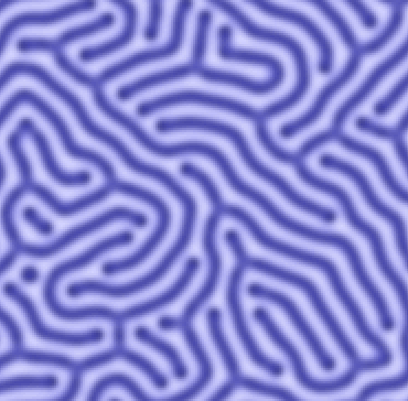
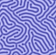 A simulation of ocular dominance column column pattern
They are enervated by input from the lateral geniculate nucleus (LGN) into layer 4 and have mostly reciprocal projections to many other parts of the visual cortex[6].
Relation to Other features of V1
The ocular dominance columns are stripe shaped and cover the primary (striate) visual cortex. If the columns corresponding to one eye were colored, a pattern similar to that shown in the accompanying figure would be visible when looking at the surface of the cortex. However, the same region of cortex could also be colored by the direction of edge that it responds to, giving the orientation columns, which are laid out in a characteristic pinwheel shape.[note 1] Similarly, there are columns in the cortex that have high levels of the protein cytochrome oxidase. These are called cytochrome oxidase "blobs" because of their scattered blob-like appearance.
All three types of column are present in the human visual cortex[3], and macaques [5]. In macaques, it was found that both blobs and pinwheel centers tend to lie in the center of ocular dominance columns.[5] No particular relation between has been found between pinwheel centers and blobs[5].
Most early models of the columns supposed that there were discrete "modules" or "hypercolumns" tiling the cortex, consisting of a repeating unit containing a full set of orientation and ocular dominance. While such units can be constructed, the map of columns is so distorted that there is no repeating structure and no clear boundaries between modules[5]. Additionally, practically every combination of having or not having orientation, dominance, and cytochrome oxidase columns has been observed in one species or another[3]. Further confusing the issue, squirrel monkeys don't always express columns, and even when they do the cytochrome oxidase blobs are not in register with the ocular dominance columns.[7]
Development
Formation
There is no consensus yet as to how ocular dominance columns are initially developed. One possibility is that they develop through Hebbian learning triggered by spontaneous activity in the eyes of the developing fetus, or in the LGN. Another possibility is that axonal guidance cues may guide the formation, or a combination of mechanisms may be at work. It is known that ocular dominance columns develop before birth, which indicates that if an activity dependent mechanism is involved it must work based on intrinsic activity.[8] It is known that spontaneous waves of activity in the retina occur before birth and that these waves are crucial for eye specific segregation of in puts to the LGN.[9] Similarly, the correlated activation for the retinal waves may direct development of the ocular dominance columns.[10] Similar spontaneous activity in the cortex may also play a role.[11][10] In any case, it has been shown that disrupting the retinal waves at least alters the pattern of ocular dominance columns.[10]
Plasticity
Sensitive periods
Although the ocular dominance columns are formed before birth, there is a period after birth--formerly called a "critical period" and now called a "sensitive period"--when the the ocular dominance columns may be modified by activity dependent plasticity. This plasticity is so strong that if the signals from both eyes are blocked the ocular dominance columns will completely desegregate[12]. Similarly, if one eye is closed ("monocular deprivation") [2],removed[13], or silenced [14] during the critical period, the size of the columns corresponding to the removed eye shrink dramatically.
Models
Many models have been proposed to explain the development and plasticity of the ocular dominance columns. In general these models can be split into two categories, those that posit formation via chemotaxis and those that posit a Hebbian activity dependent mechanism[10]. Generally chemotaxis models assume activity independent formation via the action of axon guidance molecules with the structures only later being refined by activity, but there are now known to be activity dependent [15][16] and activity modifying [17][18]guidance molecules.
Modified Hebbian learning
One major model of the formation of the stripes seen in ocular dominance columns is that they form by Hebbian competition between axon terminals.[19] The ocular dominance columns look like Turing patterns, which can be formed by modified Hebbian mechanisms. In a normal Hebbian model, if two neurons are connected to a neuron and fire together, they increase the strength of the synapses, moving the axon terminals closer together. The model must be modified to incorporate incoming activity that is locally excitatory and long range inhibitory, because if this is not done then the column width will only be dependent on the width of the axonal arbor, and also segregation will often fail in the presence of inter eye correlation. [19] This basic model has since been extended to be more physiologically plausible with the addition of long term potentiation and depression, synaptic normalization[20], neurotrophin release[21], reuptake [22], and STDP. [23]
Chemotaxis
Chemotactic models posit the existence of axon guidance molecules that direct the initial formation of the ocular dominance columns. These molecules would guide the axons as the develop based on markers specific to the axons from each eye. [10]All chemotactic models must take into account the activity dependent effects demonstrated in later development[24], but they have been called for because several pieces of evidence make entirely activity dependent formation unlikely. First, It has been shown that the ocular dominance columns in squirrel monkeys have mirror symmetry across the cortex. This is very unlikely to occur by activity dependent means because it implies a correlation between the nasal retina of one eye and the temporal retina of the other, which has not been observed. Furthermore, work in achiasmatic Belgian sheepdogs has shown that columns can form between the projections form the temporal and nasal retina of the same eye, clearly suggesting a nasal-temporal labeling, rather than contralateral vs. ipsilateral, which would be much easier to explain with activity dependent mechanisms.[25] Despite this, a molecular label that directs the formation of the ocular dominance columns has never been found. [10]
Function
It was has long been believed that ocular dominance columns play some role in binocular vision[10]. Another candidate function for ocular dominance columns (and for columns in general) is the minimization of connection lengths and processing time, which could be evolutionarily important.[26] Many believe that the ocular dominance columns serve no function[3]
Notes
- ^ A very good analogy for this is the idea of coloring a map. Just like a map of Asia could be colored by religion or by language, the columns are not physical things but regions defined by shared attributes. Also much like a map of religion the borders tend to be fuzzy with no clear distinction between one area and the next columns often don't have sharp borders. Similarly, there may be overlap, just as people at the border between France and Germany are a mixture of French speakers, German speakers, or bilingual. There are even occasional neurons belonging to the ipsilateral eye in a contralateral column just like the occasional Portuguese speaker may be found in China. It was once believed the columns were discrete units with sharp borders but the idea of fuzzy, mostly continuous regions is now preferred.[3]
References
- ^ Kolb & Whishaw: Fundamentals of Human Neuropsychology, 2003
- ^ a b http://jp.physoc.org/content/281/1/267.full.pdf#page=1&view=FitH Shatz, C. J. & Stryker, M. P. (1978) Ocular dominance in layer IV of the cat’s visual cortex and the effects of monocular deprivation. Journal of Physiology 281:267–83.
- ^ a b c d e http://hebb.mit.edu/courses/connectomics/Horton%20Adams%20cortical%20column%20structure%20without%20function%2005.pdf Horton J.C. and Adams D.L. (2005) The cortical column: a structure without a function. Phil. Trans. R. Soc. B, 360: 837-862.
- ^ Horton J.C. and Hocking D.R. (1996) Intrinsic variability of ocular dominance column periodicity in normal macaque monkeys. J. Neurosci., 16:7228-7339.
- ^ a b c d e http://www.ncbi.nlm.nih.gov/pmc/articles/PMC50666/pdf/pnas01098-0266.pdf Bartfeld E, Grinvald A. Relationships between orientation-preference pinwheels, cytochrome oxidase blobs, and ocular-dominance columns in primate striate cortex. Proc Natl Acad Sci USA. 1992;89:11905–11909.
- ^ Van Essen DC, Anderson CH, Felleman DJ. Information processing in the primate visual system: an integrated systems perspective. Science 255: 419–423, 1992.
- ^ http://www.nature.com/?file=/neuro/journal/v6/n2/full/nn1004.html&filetype=pdf Capricious expression of cortical columns in the primate brain
- ^ Crowley J.C. and Katz L.C. (2000) Early development of ocular dominance columns. Science, 290: 1321 – 1324.
- ^ http://library.ibp.ac.cn/html/cogsci/neuron-2002-357.pdf Stellwagen D, Shatz CJ. An instructive role for retinal waves in the development of retinogeniculate connectivity. Neuron 33: 357–367, 2002.
- ^ a b c d e f g http://www.ncbi.nlm.nih.gov/pmc/articles/PMC2655105/ Huberman, A.D. et al. (2008) Mechanisms underlying development of visual maps and receptive fields. Annu Rev. Neurosci. 31, 479–509
- ^ Chiu C. and Weliky M. (2002) Relationship of correlated spontaneous activity to functional ocular dominance columns in the developing visual cortex. Neuron, 35: 1123 – 1134.
- ^ http://www.jneurosci.org/content/6/8/2117.full.pdf+html Binocular Impulse Blockade Prevents the Formation of Ocular Dominance Columns in Cat Visual Cortex
- ^ Horton JC, Hocking DR. 1998. Effect of early monocular enucleation upon ocular dominance columns and cytochrome oxidase activity in monkey and human visual cortex. Vis. Neurosci. 15:289–303
- ^ http://www.keck.ucsf.edu/~idl/CV/Chapman_Oculardominance_Nature_1986.pdf Chapman, B., Jacobson, M.D., Reiter, H.O., and Stryker, M.P. (1986). Ocular dominance shift in kitten visual cortex caused by imbalance in retinal electrical activity. Nature 324, 154–156.
- ^ Hanson MG, Landmesser LT. Normal patterns of spontaneous activity are required for correct motor axon guidance and the expression of specific guidance molecules. Neuron. 2004;43:687–701.
- ^ Song HJ, Poo MM. 1999. Signal transduc- tionunderlyinggrowthconeguidanceby diffusible factors. Curr. Opin. Neurobiol. 9:355–63
- ^ Bouzioukh F, Daoudal G, Falk J, Debanne D, Rougon G, Castellani V. Semaphorin3A regulates synaptic function of differentiated hippocampal neurons. Eur. J. Neurosci. 2006;23:2247–2254.
- ^ Sahay A, et al. Secreted semaphorins modulate synaptic transmission in the adult hippocampus. J Neurosci. 2005;25:3613–3620.
- ^ a b Miller K.D., Keller J.B. and Stryker C.D. (1989) Ocular dominance column development:analysis and simulation. Science, 111:123-145.
- ^ Miller K.D (1996) Synaptic economics: competition and cooperation in correlation-based synaptic competition. Neuron, 17:371-374.
- ^ Harris A.E., Ermentrout G.B. and Small S.L. (1997) A model of ocular dominance column development by competition for trophic factor. Proc. Natl. Acad. Sci. USA, 94:9944-9949.
- ^ Elliott T. and Shadbolt N.R. (1998) Competition for neurotrophic factors: mathematical analysis. Neural computation, 10:1939-1981.
- ^ Hensch T.K. (2005) Critical period plasticity in local cortical circuits. Nat. Rev. Neurosci., 6:877-888.
- ^ Crair M.C., Horton J.C., Antonini A. and Stryker M.P. (2001) Emergence of ocular dominance columns in cat visual cortex by 2 weeks of age. J. Comparative Neurol., 430: 235-249.
- ^ Dell’Osso LF, Williams RW. Ocular motor abnormalities in achiasmatic mutant Belgian sheepdogs: unyoked eye move- ments in a mammal. Vis Res 1995;35:109–16.
- ^ http://www.nervana.montana.edu/~alex/public/constraints/Chklovskii-ocular_dominance.pdf Chklovskii DB, Koulakov AA. 2000. A wire length minimization approach to ocular dom- inance patterns in mammalian visual cortex. Physica A 284:318–34
Further Reading
- Katz LC, Crowley JC. (2002). Development of cortical circuits: lessons from ocular dominance columns. Nat Rev Neurosci, 3(1):34-42.
- Carreira-Perpinan M,Lister R, Goodhill G. 2005.A computational model for the development of multiple maps in primary visual cortex. Cerebral Cortex 15:1222–1233.
See also
- Visual cortex
- Ocular dominance
- Orientation columns
- Amblyopia
External Links
Categories:
Wikimedia Foundation. 2010.