- Orthopantomogram
-
Orthopantomogram Diagnostics MeSH D011862 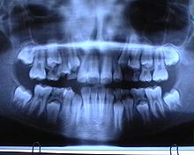 Orthopantomogram: Mixed dentition
 An OPG-Unit
An Orthopantomogram (OPT) or Dental Panoramic Radiograph (DPR), also known as an "orthopantogram" or "panorex", is a panoramic scanning dental X-ray of the upper and lower jaw. It shows a two-dimensional view of a half-circle from ear to ear. An OPT relies on tomography i.e. images of specific radiographic planes are taken to make up the larger panoramic image.
Contents
Equipment
Dental panoramic radiography equipment consists of a horizontal rotating arm which holds an X-ray source and a moving film mechanism (carrying a film) arranged at opposed extremities. The patient's skull sits between the X-ray generator and the film. The X-ray source is collimated toward the film, to give a beam shaped as a vertical blade having a width of 4-7mm[citation needed] when arriving on the film, after crossing the patient's skull. Also the height of that beam covers the mandibles and the maxilla regions. The arm moves and its movement may be described as a rotation around an instant center which shifts on a dedicated trajectory.
The manufacturers propose different solutions for moving the arm, trying to maintain constant distance between the teeth to the film and generator. Also those moving solutions try to project the teeth arch as orthogonally as possible. It is impossible to select an ideal movement as the anatomy varies very much from person to person. Finally a compromise is selected by each manufacturer and results in magnification factors which vary strongly along the film (15%-30%). The patient positioning is very critical in regard to both sharpness and distortions.
Forming the image
Normally, the person bites on a plastic spatula so that all the teeth, especially the crowns, can be viewed individually. The whole orthopantomogram process takes about one minute. The patient's actual radiation exposure time varies between 5.5 and 22 seconds for the machine’s excursion around the skull.
The collimation of the machine means that, while rotating, the X-rays project only a limited portion of the anatomy onto the film at any given instant but, as the rotation progresses around the skull, a composite picture of the maxillo-facial block is created. While the arm rotates, the film moves in a such way that the projected partial skull image (limited by the beam section) scrolls over it and exposes it entirely. Not all of the overlapping individual images projected on the film have the same magnification because the beam is divergent and the images have differing focus points. Also not all the element images move with the same velocity on the target film as some of them are more distant from and others closer to the instant rotation center. The velocity of the film is controlled in such fashion to fit exactly the velocity of projection of the anatomical elements of the dental arch side which is closest to the film. Therefore they are recorded sharply while the elements in different places are recorded blurred as they scroll at different velocity.
The dental panoramic image suffers from important distortions because a vertical zoom and a horizontal zoom both vary differently along the image. The vertical and horizontal zooms are determined by the relative position of the recorded element versus film and generator. Features closer to the generator receive more vertical zoom. The horizontal zoom is also dependent on the relative position of the element to the focal path. Features inside the focal path arch receive more horizontal zoom and are blurred; features outside receive less horizontal zoom and are blurred.
The result is an image showing sharply the section along the mandible arch, and blurred elsewhere. For example, the more radio-opaque anatomical region, the cervical vertebrae (neck), shows as a wide and blurred vertical pillar overlapping the front teeth. The path where the anatomical elements are recorded sharply is called "focal path".
Films
There are two kinds of film moving mechanisms, one using a sliding flat cassette which holds the film, and another using a rotating cylinder around which the film is wound. There are two standard sizes for dental panoramic films: 30 cm x12cm (12"x 5") and 30 cm x 15 cm (12"x6"). The smaller size film receives 8% less X-ray dosage on it compared to the bigger size.
Digital
Dental X-rays' radiology is moving from film technology (involving a chemical developing process) to Digital X-ray technology, which is based on electronic sensors and computers. One of the principal advantages compared to film based systems is the much greater exposure latitude. This means many fewer repeated scans, which reduces costs and also reduces patient exposure to radiation. Lost X-rays can also be reprinted if the digital file is saved. Other significant advantages include instantly viewable images, the ability to enhance images, the ability to email images to practitioners and clients (without needing to digitize them first), easy and reliable document handling, reduced X-ray exposure, that no darkroom is required, and that no chemicals are used.
One particular type of digital system uses a Photostimulable Phosphor Plate (aka PSP - Phosphor Plate) in place of the film. After X-ray exposure the plate (sheet) is placed in a special scanner where the latent formed image is retrieved point by point and digitized, using a laser light scanning. The digitized images are stored and displayed on the computer screen. This method is in between old film based technology and the current direct digital imaging technology. It is similar to the film process because it involves the same image support handling and differs because the chemical development process is replaced by the scanning process. This is not much faster that film processing and the resolution and sensitivity performances are contested. However it has the clear advantage of being able to fit with any existing equipment without any modification because it replaces just the existing film.
Also some times the term "Digital X-rays" is used to designate the scanned film documents which further are handled by computers.
The other types of digital imaging technologies use electronic sensors. A majority of them first convert the X-rays in light (using a GdO2S or CsI layer) which is further captured using a CCD or a CMOS image sensor. Few of them use a hybrid analog-to-digital arrangement which first converts the X-ray into electricity (using a CdTe layer) and then this electricity is rendered as an image by a reading section based on CMOS technology.
In current state-of-the-art digital systems, the image quality is vastly superior to conventional film-based systems.
Historical milestones for Digital Panoramic Systems
1985-1991 - The first dental digital panoramic systems were designed by McDavid et al. at UTHSCSA.[1]
1995 - DXIS, the first dental digital panoramic X-rays system available on the market, was introduced by Signet (France). DXIS was targeted to retrofit all the panoramic models.
1997 - SIDEXIS, of Siemens (currently Sirona Dental Systems, Germany) offered a digital option for Ortophos Plus panoramic unit, DigiPan of Trophy [1] Radiology (France) offered a digital option for the OP100 panoramic made by Instrumentarium (Finland).
1998-2004 - many panoramic manufacturers offered their own digital systems.2006 - SCAN300FP, of 'Ajat' (Finland) is the latest innovation offered. It shows the feature to acquire many hundreds of mega bytes of image information at high frame rate and to reconstruct the panoramic layer by post acquisition computing.[citation needed]
Principal advantage of panaromic images
- Broad coverage of facial bones and teeth
- Low patient radiation dose
- Convenience of examination for the patient (films need not be placed inside the mouth)
- Ability to be used in patients who cannot open the mouth or when the opening is restricted eg: due to trismus
- Short time require for making the image
- Patient's ready understandability of panaromic films, making them a useful visual aid in patient education and case presentation.
- Easy to store compared to the large set of intra oral x-rays which are typically used. [2]
Diagnostic uses
OPGs are used by Dentists to provide information on:
- Impacted wisdom teeth Diagnosis and treatment planning
- Periodontal bone loss and periapical involvement.
- Finding the source of dental pain
- Assessment for the placement of dental implants
- Orthodontic assessment. pre and post operative
- Caries detection especially in the inter-dental region.
- Diagnosis of developmental anomalies such as Cherubism, Cleido cranial dysplasia
- Carcinoma in relation to the jaws
- Tempero mandibular joint dysfuctions and ankylosis.
- Diagnosis of osteosarcoma, ameloblastoma, renal osteodystrophy affecting jaws, hypophosphatemia,
The most common use is to determine the status of wisdom teeth and trauma to the jaws.
See also
References
- ^ http://www.patentstorm.us/patents/5018177.html
- ^ Stuart C. White, Micheal J Pharoh, Oral Radiology and Interpretation Mosby 2005
Categories:


Wikimedia Foundation. 2010.