- Sphenoidal sinuses
Infobox Bone
Name = Sphenoidal sinuses
Latin = sinus sphenoidalis
GraySubject = 223
GrayPage = 998
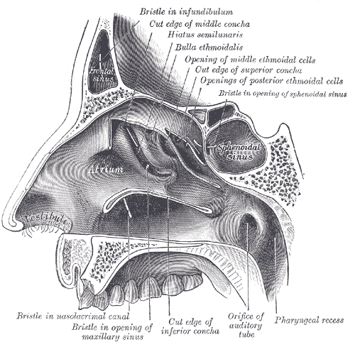
Caption = Lateral wall of nasal cavity; the three nasal conchæ have been removed. (Sphenoidal sinus visible at upper right, in dark circle.)
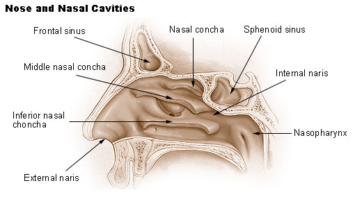
Caption2 = Nose and nasal cavities. (Sphenoid sinus labeled at upper right.)
Precursor =
System =
Artery =
Vein =
Nerve =posterior ethmoidal nerve s, and orbital branches of thepterygopalatine ganglion
Lymph =
MeshName = Sphenoid+Sinus
MeshNumber = A04.531.621.827
DorlandsPre = s_12
DorlandsSuf = 12739248
The sphenoidal sinuses (or sphenoid) contained within the body of the sphenoid vary in size and shape; owing to the lateral displacement of the interveningseptum they are rarely symmetrical.The following are their average measurements: vertical height, 2.2 cm.; transverse breadth, 2 cm.; antero-posterior depth, 2.2 cm.
Relations
When exceptionally large they may extend into the roots of the
pterygoid processes or great wings, and may invade thebasilar part of theoccipital bone .Each sinus opens into the roof of the nasal cavity via apertures on the posterior wall of the sphenoethmoidal recess. The apertures are located high on the anterior walls of the sinuses themselves.
Because only thin shelves of bone separate the sphenoidal sinuses from the nasal cavities below and hypophyseal fossa above, the pituitary gland can be surgically approached through the roof of the nasal cavities by first passing through the anterioinferior aspect of the sphenoid bone and into the sinuses, followed by entry through the top of the sphenoid bone into the hypophyseal fossa.
If a fast-growing tumor erodes the floor of the sinus, the
vidian nerve could be in danger.If the tumor spreads laterally, thecavernous sinus and all its constituent nerves could be in danger.Innervation
The mucous membrane is innervated by the
posterior ethmoidal nerve s, and the orbital branches of thepterygopalatine ganglion .Infection
A potential complication of sphenoid sinusitis is cavernous sinus thrombosis.
Development
They are present as minute cavities at birth, but their main development takes place after
puberty .
=Additionalee also
*
Paranasal sinus External links
*
* (NormanAnatomyFig|latnasalwall3)
Wikimedia Foundation. 2010.