- Cohesin
-
This article is about the protein complex. For the protein domain in cellulose degrading bacteria, see Cohesin domain.
Sister chromatid cohesion protein 1 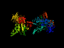
Cohesin complex composed of
SCC1 and SMC1 proteins.[1]Identifiers Symbol MCD1 Alt. symbols PDS3, RHC21, SCC1 Entrez 851561 PDB 1W1W UniProt Q12158 Other data Cohesin subunit SCC3 Identifiers Symbol IRR1 Alt. symbols SCC3 Entrez 854786 UniProt P40541 Other data Structural maintenance of chromosomes protein 1 Identifiers Symbol SMC1 Alt. symbols CHL10 Entrez 850540 PDB 1W1W UniProt P32908 Other data Structural maintenance of chromosomes protein 3 Identifiers Symbol SMC3 Alt. symbols J1049 Entrez 853371 UniProt P47037 Other data Cohesin is a protein complex that regulates the separation of sister chromatids during cell division, either mitosis or meiosis.
Contents
Structure
Cohesin is made up of four subunits, Scc1, Scc3, Smc1 and Smc3. Smc1 and Smc3 are members of the Structural Maintenance of Chromosomes (SMC) family. SMC proteins have two main structural characteristics: an ATPase activity domain (formed when the amino and carboxy terminals interact) and a hinge region that allows dimerization of SMCs. The ATPase domain and the hinge are connected to each other via long anti-parallel coiled coils. The overall structure of a dimer has, therefore, an ATPase domain at each end and a hinge at the center. When ATP is present, the two ATPase domains are able to bind, forming a ring structure. ATP hydrolysis can therefore trigger opening and closing of the ring.[2]
Scc1 and Scc3 bind the ATPase domains of Smc1 and Smc3 stabilizing the ring structure. Scc1 is a member of the kleisin protein family and it controls sister-chromatid separation. The amino and carboxy terminus of Scc1 bind Smc1 and Smc3. Once Scc1 binds on the SMC proteins, Scc3 can also associate by binding with the C-terminal region of Scc1. When Scc1 binds on both Smc1 and Smc3, the cohesin complex forms a closed ring structure. When it binds to only one of the SMC proteins, the complex forms an open ring.[3] However more recently cohesin rings were found to dimerise, with two rings held together by the Scc3 subunit in a handcuff shape, one strand of DNA in each cohesin ring. [4]
Function
The cohesin ring has three functions:
1. It is used to keep the sister chromatids connected with each other during metaphase ensuring that during mitosis (and meiosis), each sister chromatid segregates to opposite poles. Without cohesin, the cell would be unable to control sister chromatid segregation since there would be no way of ensuring whether the spindle fiber attached on each sister chromatid is from a different pole.
2. It facilitates spindle attachment onto chromosomes.
3. It facilitates DNA repair by recombination.
Dissociation of sister chromatid cohesion
The anaphase promoting complex associated to Cdc20 (APC/C-cdc20) cleaves the securin (anaphase inhibitor), which holds the sister chromatids together. Securin is cleaved at anaphase, following APC/C-cdc20 mediated degradation, and it renders separase (a protease, inhibited by the association with securin) to cleave kleisin. Kleisin is associated with the cohesin complex, linking both SMC 3 and SMC 1 together, ultimately accomplishing the proteolysis of cohesin.
Dissociation of sister chromatids cohesion defines anaphase onset, which establishes two sets of identical chromosomes at each pole of the cell (telophase). Then the two daughter cells separate, and a new round of the cell cycle freshly starts in each one, at the stage of G0. When cells are ready to divide, because cell size is big enough or because they receive the appropriate stimulus,[5] they activate the mechanism to enter into the G1 stage of cell cycle, and they duplicate most organelles during S (synthesis) phase, including their centrosome. Therefore, when the cell division process will end, each daughter cell will receive a complete set of organelles. At the same time, during S phase all cells must duplicate their DNA very precisely, a process termed DNA replication. Once DNA replication has finished, in eukaryotes the DNA molecule is compacted and condensed, to form the mitotic chromosomes, each one constituted by two sister chromatids, which stay hold together by the establishment of cohesion between them; each chromatid is a complete DNA molecule, attached via microtubules to one of the two centrosomes of the dividing cell, located at opposed poles of the cell. To avoid premature sister chromatid separation, the APC/C is maintained in an inactive state bound to different molecules, which are part of a complex mechanism termed the spindle assembly checkpoint.
Mechanism of action
It is not clear how the cohesion ring links sister chromatids together. There are two possible scenarios:
- Cohesin subunits bind to each sister chromatid and form a bridge between the two.
- Since cohesin has a ring structure, it is able to encircle both sister chromatids.
Current evidence suggests that the second scenario is the most likely. Proteins that are essential for sister chromatid cohesion, such as Smc3 and Scc1, do not regulate the formation of covalent bonds between cohesin and DNA, indicating that DNA interaction is not sufficient for cohesion.[3] In addition, disturbing the ring structure of cohesin through cleavage of Smc3 or Scc1 triggers premature sister chromatid segregation in vivo.[6] This shows that the ring structure is important for cohesin’s function.
Even though the ring hypothesis appears to be valid, there are still questions about the number of rings required to hold sister chromatids together. One possibility is that one ring surrounds the two chromatids. Another possibility involves the creation of a dimer where each ring surrounds one sister chromatid. The two rings are connected to each other through formation of a bridge that holds the two sister chromatids together.
The cohesion complex is established during the initial stages of S-phase. The complexes associate with chromosomes before DNA replication occurs. Once cells start replicating their DNA, cohesin rings close and link the sister chromatids together.[3] Cohesin complexes must be present during S-phase in order for cohesion to take place. It is unclear, however, how cohesin is loaded on the chromosomes during G1. There are two proposed hypotheses so far:
- The ATPase domain of the SMC proteins interacts with DNA and this interaction initially mediates the loading of cohesin complexes on chromosomes.
- Several proteins aid in the loading process. For example, Scc2 and Scc4 are both required for cohesin to load in budding yeast.
Localization of cohesin rings
Cohesin binding along the chromosomal DNA is considered to be dynamic and its location changes based on gene transcription, specific DNA sequence and presence of chromosome-associated proteins. There are three possible scenarios:
- Cohesin location is influenced by the orientation of neighboring genes and it is most frequently located in areas of convergent transcription. Gene orientation depends on the direction of transcription and can be of three types: head-to-head, head-to-tail and tail-to-tail. The tail-to-tail configuration results in the convergence of transcription machinery. One hypothesis states that the RNA polymerase “pushes” cohesin along the DNA, causing them to move towards the direction of the RNA polymerases. Changing the transcription pattern of genes changes the location of cohesin indicating that localization of cohesin may depend on transcription.[7]
- A few cohesin rings are found in chromosome arms that have AT-rich DNA sequences indicating that DNA sequence may be an independent factor of cohesin binding.[7]
- Cohesin rings, especially in budding yeast, are also located in the region surrounding the centromere.[7] Two hypotheses may explain this: the presence of repetitive heterochromatic DNA in centromeres and the presence of chromosome-associated proteins. For example, Schizosaccharomyces pombe have multiple copies of specific heterochromatic DNA whose involvement in cohesion binding has been proven. Budding yeast lacks repetitive sequences and, therefore, requires a different mechanism for cohesion binding. Evidence suggests that binding of cohesin to the budding yeast centromere region depends on chromosome-associated proteins of the kinetochore that mediate cohesion association to pericentric regions (the kinetochore is an enhancer of pericentric cohesin binding).[8]
Evolution
Cohesin structure and function has been conserved in evolution. The SMC proteins are found in prokaryotes and have been conserved through evolution. The coiled coils of SMC1 and SMC3 are conserved with an amino acid divergence of less than 0.5%.[9]
Name Saccharomyces Cerevisiae Schizosaccharomyces pombe Drosophila Vertebrates Smc1 Smc1 Psm1 DmSmc1 Smc1 Smc3 Smc3 Psm3 DmSmc3 Smc3 Scc1 Mcd1/Pds3 Rad21 DmRad21 Rad21 Scc3 Scc3 Psc3 DmSA SA1 and SA2 Clinical significance
The term "cohesinopathy" has been used to describe conditions affecting the cohesin complex.[10][11][12]
These conditions include:
- Cornelia de Lange Syndrome
- Roberts syndrome
- Warsaw breakage syndrome
See also
- Condensin
- SMC protein
- Establishment of sister chromatid cohesion
References
- ^ PDB 1W1W; Haering CH, Schoffnegger D, Nishino T, Helmhart W, Nasmyth K, Löwe J (September 2004). "Structure and stability of cohesin's Smc1-kleisin interaction". Mol. Cell 15 (6): 951–64. doi:10.1016/j.molcel.2004.08.030. PMID 15383284.
- ^ Morgan DL (2007). The cell cycle: principles of control. London: Published by New Science Press in association with Oxford University Press. ISBN 0-87893-508-8.
- ^ a b c Gruber S, Haering CH, Nasmyth K (March 2003). "Chromosomal cohesin forms a ring". Cell 112 (6): 765–77. doi:10.1016/S0092-8674(03)00162-4. PMID 12654244.
- ^ Nenggang Zhang, Sergey G. Kuznetsov, Shyam K. Sharan, Kaiyi Li1, Pulivarthi H. Rao1, and Debananda Pati. A handcuff model for the cohesin complex. Journal of Cell Biology. Vol. 183: 1019-1031 [1]
- ^ Conlon, Ian; Raff, Martin (1999). "Size Control in Animal Development". Cell 96 (2): 235–44. doi:10.1016/S0092-8674(00)80563-2. PMID 9988218.
- ^ Peters JM, Tedeschi A, Schmitz J (November 2008). "The cohesin complex and its roles in chromosome biology". Genes Dev. 22 (22): 3089–114. doi:10.1101/gad.1724308. PMID 19056890.
- ^ a b c Ross KE, Cohen-Fix O (July 2004). "Molecular biology: cohesins slip sliding away". Nature 430 (6999): 520–1. doi:10.1038/430520b. PMID 15282594.
- ^ Weber SA, Gerton JL, Polancic JE, DeRisi JL, Koshland D, Megee PC (September 2004). "The kinetochore is an enhancer of pericentric cohesin binding". PLoS Biol. 2 (9): E260. doi:10.1371/journal.pbio.0020260. PMC 490027. PMID 15309047. http://www.pubmedcentral.nih.gov/articlerender.fcgi?tool=pmcentrez&artid=490027.
- ^ White GE, Erickson HP (2009). "The coiled coils of cohesin are conserved in animals, but not in yeast". PLoS ONE 4 (3): e4674. doi:10.1371/journal.pone.0004674. PMC 2650401. PMID 19262687. http://www.pubmedcentral.nih.gov/articlerender.fcgi?tool=pmcentrez&artid=2650401.
- ^ Gard S, Light W, Xiong B, et al. (November 2009). "Cohesinopathy mutations disrupt the subnuclear organization of chromatin". J. Cell Biol. 187 (4): 455–62. doi:10.1083/jcb.200906075. PMC 2779225. PMID 19948494. http://www.pubmedcentral.nih.gov/articlerender.fcgi?tool=pmcentrez&artid=2779225.
- ^ van der Lelij P, Chrzanowska KH, Godthelp BC, et al. (February 2010). "Warsaw breakage syndrome, a cohesinopathy associated with mutations in the XPD helicase family member DDX11/ChlR1". Am. J. Hum. Genet. 86 (2): 262–6. doi:10.1016/j.ajhg.2010.01.008. PMC 2820174. PMID 20137776. http://www.pubmedcentral.nih.gov/articlerender.fcgi?tool=pmcentrez&artid=2820174.
- ^ van der Lelij P, Godthelp BC, van Zon W, et al. (2009). "The cellular phenotype of Roberts syndrome fibroblasts as revealed by ectopic expression of ESCO2". PLoS ONE 4 (9): e6936. doi:10.1371/journal.pone.0006936. PMC 2734174. PMID 19738907. http://www.pubmedcentral.nih.gov/articlerender.fcgi?tool=pmcentrez&artid=2734174.
Further reading
- Michaelis C, Ciosk R, Nasmyth K (October 1997). "Cohesins: chromosomal proteins that prevent premature separation of sister chromatids". Cell 91 (1): 35–45. doi:10.1016/S0092-8674(01)80007-6. PMID 9335333.
- Guacci V, Koshland D, Strunnikov A (October 1997). "A direct link between sister chromatid cohesion and chromosome condensation revealed through the analysis of MCD1 in S. cerevisiae". Cell 91 (1): 47–57. doi:10.1016/S0092-8674(01)80008-8. PMC 2670185. PMID 9335334. http://www.pubmedcentral.nih.gov/articlerender.fcgi?tool=pmcentrez&artid=2670185.
- Tóth A, Ciosk R, Uhlmann F, Galova M, Schleiffer A, Nasmyth K (February 1999). "Yeast cohesin complex requires a conserved protein, Eco1p(Ctf7), to establish cohesion between sister chromatids during DNA replication". Genes Dev. 13 (3): 320–33. doi:10.1101/gad.13.3.320. PMC 316435. PMID 9990856. http://www.pubmedcentral.nih.gov/articlerender.fcgi?tool=pmcentrez&artid=316435.
- Uhlmann F, Lottspeich F, Nasmyth K (July 1999). "Sister-chromatid separation at anaphase onset is promoted by cleavage of the cohesin subunit Scc1". Nature 400 (6739): 37–42. doi:10.1038/21831. PMID 10403247.
External links
Categories:- Mitosis
- Proteins

Wikimedia Foundation. 2010.