Tuberosity of the ischium
- Tuberosity of the ischium
Infobox Bone
Name = Tuberosity of the ischium
Latin = tuber ischiadicum
GraySubject = 57
GrayPage = 235
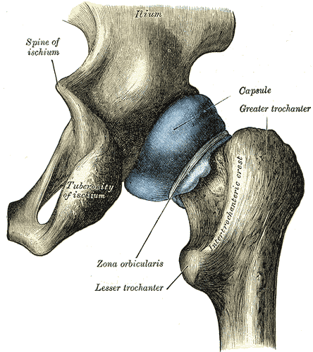
Caption = Capsule of hip-joint (distended). Posterior aspect. (Tuberosity of ischium visible at bottom left.)
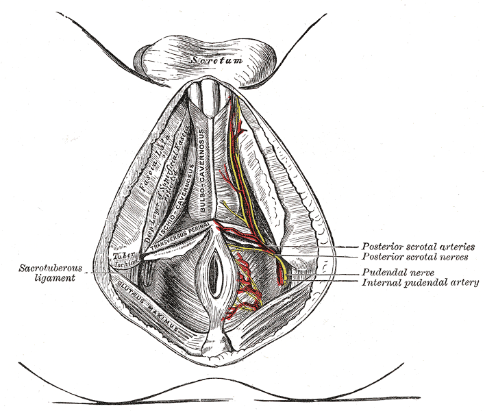
Caption2 = The superficial branches of the internal pudendal artery. (Tuber. ischial. visible at center left.)
System =
MeshName =
MeshNumber =
DorlandsPre = t_21
DorlandsSuf = 12827506
Posteriorly, the superior ramus of the ischium forms a large swelling, the tuberosity of the ischium (or ischial tuberosity).
It marks the lateral boundary of the pelvic outlet.
When sitting, the weight is frequently placed upon the ischial tuberosity. [cite journal | author = Goossens R, Teeuw R, Snijders C | title = Sensitivity for pressure difference on the ischial tuberosity. | journal = Ergonomics | volume = 48 | issue = 7 | pages = 895–902 | year = 2005 | pmid = 16076744 | doi = 10.1080/00140130500123647]
Divisions
The tuberosity is divided into two portions: a lower, rough, somewhat triangular part, and an upper, smooth, quadrilateral portion.
* The "lower portion" is subdivided by a prominent longitudinal ridge, passing from base to apex, into two parts;
** the outer gives attachment to the Adductor magnus,
** the inner to the sacrotuberous ligament.
* The "upper portion" is subdivided into two areas by an oblique ridge, which runs downward and outward;
** from the upper and outer area the Semimembranosus arises;
** from the lower and inner, the long head of the Biceps femoris and the Semitendinosus.
ee also
* Sitting disability
=Additional
References
External links
* - "The Female Perineum: Bones"
* - "Major Joints of the Lower Extremity: Hip bone (lateral view)"
* (NormanAnatomyFig|pelvisposterior, NormanAnatomyFig|pelvislateral, NormanAnatomyFig|pelvisinside)
Wikimedia Foundation.
2010.
Look at other dictionaries:
Superior ramus of the ischium — Infobox Bone Name = Superior ramus of the ischium Latin = ramus superior ossis ischii GraySubject = 57 GrayPage = 235 Caption = Right hip bone. External surface. Caption2 = System = MeshName = MeshNumber = DorlandsPre = r 02 DorlandsSuf =… … Wikipedia
tuberosity — A large tubercle or rounded elevation, especially from the surface of a bone. SYN: tuberositas [TA]. bicipital t. SYN: radial t.. calcaneal t. [TA] the posterior extremity of the calcaneus, or os calcis, forming the … Medical dictionary
List of muscles of the human body — Skeletal muscles homo sapiens Muscles of the human body: Overview Head | Neck |&# … Wikipedia
Muscular system of the horse — See also: Equine anatomy Contents 1 Types of muscle 2 Build of skeletal muscle 3 Tendons of the lower leg 4 Main skeletal muscles of the horse … Wikipedia
ischial tuberosity — n a bony swelling on the posterior part of the superior ramus of the ischium that gives attachment to various muscles and bears the weight of the body in sitting * * * tuber ischiadicum … Medical dictionary
Posterior branch of the obturator artery — Infobox Artery Name = PAGENAME Latin = ramus posterior arteriae obturatoriae GraySubject = 155 GrayPage = 616 Caption = Caption2 = BranchFrom = obturator artery BranchTo = Vein = Supplies = MeshName = MeshNumber = DorlandsPre = r 02 DorlandsSuf … Wikipedia
pubotuberous diameter — the distance from the tuberosity of the ischium to a point on the superior ramus of the pubis which is located directly perpendicular to the tuberosity … Medical dictionary
Bone — This article is about the skeletal organ. For other uses, see Bone (disambiguation) and Bones (disambiguation). For the tissue, see Osseous tissue. Drawing of a human femur Bones are rigid organs that constitute part of the endoskeleton of… … Wikipedia
Adductor magnus muscle — The adductor magnus and nearby muscles … Wikipedia
Hip bone — Left hip joint, opened by removing the floor of the acetabulum from within the pelvis. Plan … Wikipedia