- Cingulate cortex
-
Brain: Cingulate cortex 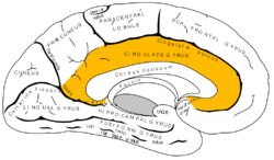
Medial surface of left cerebral hemisphere. 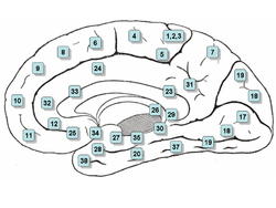
Medial surface. Latin Cortex cingularis Gray's subject #189 825 Part of Cerebral cortex Artery Anterior cerebral Vein Superior sagittal sinus Acronym(s) Cg NeuroNames hier-141 MeSH Gyrus+Cinguli NeuroLex ID birnlex_934 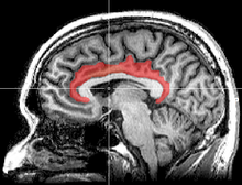 Sagittal MRI slice with highlighting indicating location of the cingulate cortex.
 Coronal section of brain. Cingulate cortex is shown in yellow.
The cingulate cortex is a part of the brain situated in the medial aspect of the cortex. It includes the cortex of the cingulate gyrus, which lies immediately above the corpus callosum, and the continuation of this in the cingulate sulcus. The cingulate cortex is usually considered part of the limbic lobe, separate from the adjacent frontal and parietal lobes.
It receives inputs from the thalamus and the neocortex, and projects to the entorhinal cortex via the cingulum. It is an integral part of the limbic system, which is involved with emotion formation and processing, learning, and memory, and is also important for executive function and respiratory control.
Contents
History
Cingulum means belt in Latin. The name was likely chosen because this cortex, in great part, surrounds the corpus callosum. Cingulate is an adjective (cingularis or cingulatus).
The cingulate cortex is a part of the "grand lobe limbique" of Broca (1898) that consisted (in addition to the olfactory part, which is no more considered there today) of a superior cingulate part, supracallosal; and an inferior hippocampic part, infracallosal. The limbic lobe was separated from the remainder of the cortex by Broca for two reasons: first that it is not convoluted, and second that the gyri are directed parasagittally (contrary to the transverse gyrification). Since the parasagittal gyrification is observed in non-primate species, the limbic lobe was thus declared to be "bestial". As with other parts of the cortex, there have been and continue to be discrepancies concerning boundaries and naming. Brodmann (1909), a student of Cécile Vogt-Mugnier and Oskar Vogt, who worked on cercopithecus (and not much in human (Bailey and von Bonin)), elaborated a system of numeration that had unfortunately no typological logics (1, 2 and 3 are sensory, 4 is motor, 5 is parietal, 6 is premotor and 7 is again parietal!). Area 25 was even not placed by him in the same place in the human brain. Area 24 (anterior) was distinguished from 23 (posterior) on the basis that it was agranular. More recently, the typographical von Economo's system was adopted by Bailey and von Bonin. Simple typographical naming should be preferred, for evident heuristic purposes.
Subdivisions
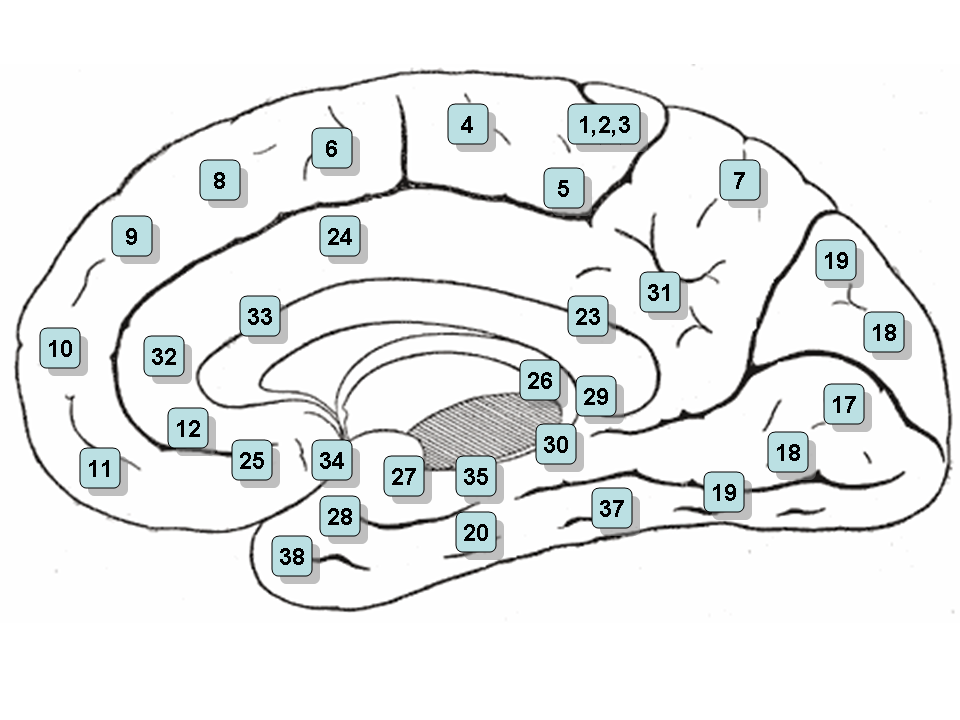
Based on cerebral cytoarchitectonics it has been divided into the Brodmann areas 23, 24, 26, 29, 30, 31 and 32. The areas 26, 29 and 30 are usually referred to as the retrosplenial areas.
Anterior cingulate cortex
Main article: Anterior cingulate cortexThis corresponds to area 24 of Brodmann and LA of Constantin von Economo and Bailey and von Bonin. It is continued anteriorly by the subgenual cortex (area 25). It is cytoarchitectonically agranular. It has a gyral part on the surface and a sulcal part. Anterior cingulate cortex can further be divided in the perigenual anterior cingulate cortex and midcingulate cortex. The anterior cingulate cortex receives primarily its afferent axons from the intralaminar and midline thalamic nuclei (intralaminar and midline of the thalamus, see thalamus). The nucleus anterior receives mamillo-thalamic afferences. The mamillary neurons receive axons from the subiculum. The whole forms a part of Papez' circuit. The anterior cingulate cortex sends axons to the anterior nucleus and through the cingulum to other Broca's limbic areas. The ACC is involved in error and conflict detection processes, such as in the go/no-go task.
Posterior cingulate cortex
Main article: Posterior cingulate cortexThis corresponds to area 23 of Brodmann LP of von Economo and Bailey and von Bonin. Its cellular structure is granular. It is followed posteriorly by the retrosplenial cortex (area 29).[citation needed] Dorsally is the granular area 31. The posterior cingulate cortex receives a great part of its afferent axons from the superficial nucleus (or nucleus superior- falsely LD-[citation needed]) of the thalamus (see thalamus), which itself receives axons from the subiculum. To some extent it thus duplicates Papez' circuit. It receives also direct afferents from the subiculum of the hippocampus.
Inputs of the Anterior Cingulate Gyrus
A retrograde tracing experiment on macaque monkeys revealed that ventral anterior (VA) and ventral lateral (VL) nuclei of the thalamus are connected with motor areas of cingulate sulcus (McFarland and Haber, 2000). Retrosplenial region (Brodmann’s area 26, 29 and 30) part of cingulate gyrus can be divided into three parts retrosplenial granular cortex A, the retrosplenial granular cortex B and the retrosplenial dysgranular cortex. The hippocampal formation sends dense projections to the retrosplenial granular cortex A and B and fewer projections to the retrosplenial dysgranular cortex. Postsubiculum sends projections to the retrosplenial granular cortex A and B and to the retrosplenial dysgranular cortex. Dorsal subiculum sends projections to the retrosplenial granular cortex B, while ventral subiculum sends projections to the retrosplenial granular cortex A. Etnorhinal cortex – caudal parts – sends projections to the retrosplenial dysgranular cortex (Wyss & Groen, 1999).
Outputs of the Anterior Cingulate Gyrus
Rostral cingulate gyrus (Brodmanns’s area 32) projects to rostral superior temporal gyrus, midorbitofrontal cortex and lateral prefrontal cortex (Pandya, Van Hoesen & Mesulam, 1981). Ventral anterior cingulate (Brodmann’s area 24) sends projections to the anterior insular cortex, premotor cortex (Brodmann’s area 6), Brodmann’s area 8, the perirhinal area, the orbitofrontal cortex (Brodmann’s area 12), the laterobasal nucleus of amygdala, and the rostral part of the inferior parietal lobule (Pandya, Van Hoesen & Mesulam, 1981). Injecting wheat germ agglutinin and horseradish peroxidase conjugate into anterior cingulate gyrus of cats, revealed that anterior cingulate gyrus has reciprocal connections with rostral part of the thalamic posterior lateral nucleus and rostral end of the pulvinar (Fuji, 1983). Postsubiculum receives projections from the retrospleinal dysgranular cortex and the retrosplenial granular cortex A and B. Parasubiculum receives projections from the retrosplenial dysgranular cortex and retrosplenial granular cortex A. Caudal and lateral parts of the entorhinal cortex get projections from the retrosplenial dysgranular cortex, while caudal medial entorhinal cortex receives projections from the retrosplenial granular cortex A. The retrosplenial dysgranular cortex sends projections to the perirhinal cortex. The retrospleinal granular cortex A sends projection to the rostral presubiculum (Wyss & Groen, 1999).
Outputs of the Posterior Cingulate Gyrus
The posterior cingulate cortex (Brodmann’s area 23) sends projections to dorsolateral prefrontal cortex (Brodmann’s area 9), anterior prefrontal cortex (Brodmann’s area 10), orbitofrontal cortex (Brodmanns’ area 11), the parahippocampal gyrus, posterior part of the inferior parietal lobule, the presubiculum, the superior temporal sulcus and the retrosplenial region (Pandya, Van Hoesen & Mesulam, 1981). The retrosplenial cortex and caudal part of the cingulate cortex are connected with rostral prefrontal cortex via cingulate fascicule in macaque monkeys (Petrides and Pandya, 2007). Ventral posterior cingulate cortex was found to be reciprocally connected with the caudal part of the posterior parietal lobe in rhesus monkeys (Cavada and Goldman-Raiuc, 1989). Also the medial posterior parietal cortex is connected with posterior ventral bank of the cingulate sulcus (Cavada and Goldman-Raiuc, 1989).
Other Connections
The anterior cingulate is connected to the posterior cingulate at least in rabbits. Posterior cingulate gyrus is connected with retrosplenial cortex and this connection is part of the dorsal splenium of the corpus callosum. The anterior and posterior cingulate gyrus and retrosplenial cortex send projections to subiculum and presubiculum (Adey, 1951).
Cingulate Gyrus and Schizophrenia
Using a three-dimensional magnetic resonance imaging procedure to measure the volume of the rostral anterior cingulate gyrus (perigenual cingulate gyrus) Takahashi et. all (2003) found that the rostral anterior cingulate gyrus is larger in control – healthy – females than males, but this sex difference was not found in schizophrenic patients. Schizophrenic patients also had a smaller volume of perigenual cingulate gyrus than control subjects. Haznedar et. all (2004) studied metabolic rate of glucose in anterior and posterior cingulate gyrus in patients with schizophrenia, schizotypal personality disorder (SPD) and compared them with a control group. The metabolic rate of glucose was found to be lower in the left anterior cingulate gyrus and the right posterior cingulate gyrus in patients with schizophrenia relative to controls. Although SPD patients were expected to show a glucose metabolic rate somewhere between the schizophrenic and controls, they actually had higher metabolic glucose rate in the left posterior cingulate gyrus ( Haznedar et all, 2004). The volume of the left anteriror cingulate gyrus was reduced in schizophrenic patients as compared with controls, but there was not any difference between SPD patients and schizophrenic patients. From these results it appears that the schizophrenia and SPD are two different disorders. A study of the volume of the gray and white matter in the anterior cingulate gyrus in patients with schizophrenia and their healthy first and second degree relatives revealed no significant difference in the volume of the white matter in the schizophrenic patients and their healthy relatives (Costain et. all, 2010). Nonethless a significant difference in the volume of gray matter was detected, schizophrenic patients had smaller volume of gray matter than their second degree relatives, but not relative to their first degree relatives. Both schizophrenic and their first degree healthy relatives have smaller gray matter volume than the second degree healthy relatives. It appears that genes are responsible for the decreased volume of gray matter in schizophrenic patients (Costain et. all, 2010). Fujiwara et. all (2007) did an experiment in which they correlated the size of anterior cingulate gyrus in schizophrenic patients with their functioning on social cognition, psychopathology and emotions with control group. The smaller the size of anterior cingulate gyrus, the lower was the level of social functioning and the higher was the psychopathology in schizophrenic patient. The anterior cingulate gyrus was found to be bilaterally smaller in patients with schizophrenia as compared with control group. No difference in IQ tests and basic visuoperceptual ability with facial stimuli was found between schizophrenia patients and the control.
Summary
Schizophrenic patients have differences in the anterior cingulate gyrus when compared with controls. Anterior cingulate gyrus was found to be smaller in schizophrenic patients (Fujiwara et. all, 2007). The volume of the white and gray matter in the anterior cingulate gyrus was found to be lower in schizophrenic patients (Haznedar et. all 2004). Healthy females have larger rostral anterior cingulate gyrus than males, this sex difference in size is absent in schizophrenic patients (Takahashi et. all, 2003). Metabolic rate of glucose was lower in the left anterior cingulate gyrus and in the right posterior cingulate gyrus (Haznedar et. all, 2004). In addition to changes in the cingulate cortex more brain structures show changes in schizophrenic patients as compared to controls. The hippocampus in schizophrenic patients was found to decrease in size when compared with controls of the same age group (Koolschijn et. all, 2010). The caudate and putamen was found to decrease in volume in a longitudinal study of schizophrenic patients (Mitelman et. all, 2009). While the volume of gray matter decreases, the size of the ventricles increases in schizophrenic patients, both lateral ventricles and third ventricle (Kempton et. all, 2010).
External links
- Four Regions of Cingulate Cortex and Disease Vulnerability, Brent A. Vogt.
- BrainMaps at UCDavis Cingulate
- Mapping 'self' and 'other' in the brain
- A nice picture of the cingulate cortex and its parts
- NIF Search - Cingulate Cortex via the Neuroscience Information Framework
References
Adey, W. R. (1951). An experimental study of the hippocampal connexions of thecingulate cortex in the rabbit. Brain (1951) 74(2): 233-24.
Cavada, C., Goldman-Raiuc, P. S. (1989). Posterior Parietal Cortex in Rhesus Monkey: I. Parcellation of Areas Based on Distinctive Limbic and Sensory Corticocortical Connections. The Journal of Comparative Neurology, 287:393-421.
Costain, G., Ho, A., Crawley, A. P., Mikulis, D. J., Brzustowicz, L. M., Chow, E. W. C., Bassett, A. S. (2010). Reduced gray matter in the anterior cingulate gyrus in familial schizophrenia: A preliminary report. Schizophrenia Research 122 (2010) 81–84. Haznedar, M. M., Buchsbaum, M. S., Hazletta, E. A., Shihabuddina, L. , Newa, A., Sievera, L. J. (2004). Cingulate gyrus volume and metabolism in the schizophrenia spectrum. Schizophrenia Research 71 (2004) 249– 262. Fujiwara, H., Hirao, K., Namiki, C., Yamada, M., Shimizu, M., Fukuyama, H., Hayashi, T., Murai, T. (2007). Anterior cingulate pathology and social cognition in schizophrenia: A study of gray matter, white matter and sulcal morphometry. NeuroImage 36 (2007) 1236–1245.
Kempton M.J., Stahl D., Williams S.C., DeLisi L.E. (2010). Progressive lateral ventricular enlargement in schizophrenia: a meta-analysis of longitudinal MRI studies. Schizophr Res. 2010 Jul;120(1-3):54-62. Epub 2010 May 26.
Koolschijn P.C., van Haren N.E., Cahn W., Schnack H.G., Janssen J., Klumpers F., Hulshoff Pol H.E., Kahn R.S. (2010). Hippocampal volume change in schizophrenia. J Clin Psychiatry. 2010 Jun;71(6):737-44. Epub 2010 Jan 12.
Masako, F. (1983). Fiber Connections Between the Thalamic Posterior Lateral Nucleus and the Cingulate Gyrus in the Cat. Neuroscience Letters, 39 (1983) 137-14.
McFarland, N. R., Harber, S. N. (2000). Convergent Inputs from Thalamic Motor Nuclei and Frontal Cortical Areas to the Dorsal Striatum in the Primate. The Journal of Neuroscience, May 15, 2000, 20(10):3798–3813.
Mitelman S.A., Canfield E.L., Chu K.W., Brickman A.M., Shihabuddin L., Hazlett E.A., Buchsbaum M.S. (2009). Poor outcome in chronic schizophrenia is associated with progressive loss of volume of the putamen. Schizophr Res. 2009 Sep;113(2-3):241-5. Epub 2009 Jul 18.
Pandya, D. N., Van Hoesen, G. W., Mesulam, M. M. (1981). Efferent Connections of the Cingulate Gyrus in the Rhesus Monkey. Experimental Brain Research (1981) 42:319-330.
Petrides, M., Pandya, D. N. (2007). Efferent Association Pathways from the Rostral Prefrontal Cortex in the Macaque Monkey. The Journal of Neuroscience, 27(43):11573–11586 • 11573.
Takahashi, T., Suzuki, M., Kawasaki, Y., Hagino, H., Yamashita, I., Nohara, S., Nakamura, K., Seto, H., Kurachi, M. (2003). Perigenual Cingulate Gyrus Volume in Patients with Schizophrenia: A Magnetic Resonance Imaging Study. Biological Psychiatry 2003;53:593–600.
Wyss, J. M., Van Groen, T. (1992). Connections Between the Retrosplenial Cortex and the Hippocampal Formation in the Rat: A Review. Hippocampus, vol. 2, no. 1, pages 1-12, January 1992.
Anxiety disorder: Obsessive–compulsive disorder (F42, 300.3) History Yale–Brown Obsessive Compulsive ScaleBiology NeuroanatomyBasal ganglia (striatum) · Orbitofrontal cortex · Cingulate cortex · Brain-derived neurotrophic factorReceptorsSymptoms Obsessions (associative, diagnostic, injurious, scrupulous, pathogenic, sexual) · Compulsions (impulses, rituals, tics) · Thought suppression (avoidance) · Hoarding (animals, books, possessions)Treatment Mu opioidergicsNMDA glutamatergicsNK-1 tachykininergicsOtherBehavioralOrganizations Notable people Edna B. Foa · Stanley Rachman · Adam S. Radomsky · Jeffrey M. Schwartz · Susan Swedo · Jeff Bell · Emily ColasPopular culture LiteratureFictionalNonfictionEverything in Its Place · Just CheckingMediaRelated Obsessive–compulsive personality disorder · Obsessional jealousy · Purely Obsessional OCD · Social anxiety disorder · Tourette syndromeCategories:


Wikimedia Foundation. 2010.