- Body of humerus
-
Bone: Body of humerus 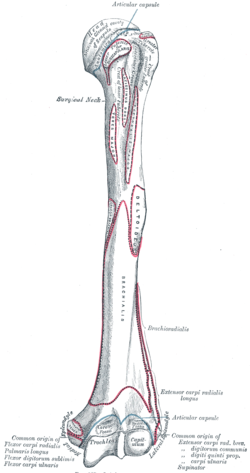
Left humerus. Anterior view. 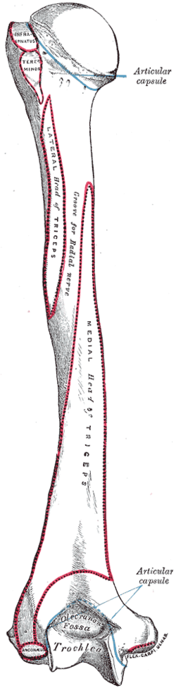
Left humerus. Posterior view. Latin corpus humeri Gray's subject #51 209 The body or shaft of the humerus is almost cylindrical in the upper half of its extent, prismatic and flattened below, and has three borders and three surfaces.
Contents
Borders
Anterior
The anterior border runs from the front of the greater tubercle above to the coronoid fossa below, separating the antero-medial from the antero-lateral surface. Its upper part is a prominent ridge, the crest of the greater tubercle; it serves for the insertion of the tendon of the pectoralis major muscle. About its center it forms the anterior boundary of the deltoid tuberosity, on which the deltoid muscle attaches; below, it is smooth and rounded, affording attachment to the brachialis muscle.
Lateral
The lateral border runs from the back part of the greater tubercle to the lateral epicondyle, and separates the anterolateral from the posterior surface. Its upper half is rounded and indistinctly marked, serving for the attachment of the lower part of the insertion of the teres minor muscle, and below this giving origin to the lateral head of the triceps brachii muscle; its center is traversed by a broad but shallow oblique depression, the spiral groove (musculospiral groove). The radial nerve runs in the spiral groove. Its lower part forms a prominent, rough margin, a little curved from backword, farword the lateral supracondylar ridge, which presents an anterior lip for the origin of the brachioradialis muscle above, and extensor carpi radialis longus muscle above, a posterior lip for the triceps brachii muscle, and an intermediate ridge for the attachment of the lateral intermuscular septum.
Medial
The medial border extends from the lesser tubercle to the medial epicondyle. Its upper third consists of a prominent ridge, the crest of the lesser tubercle, which gives insertion to the tendon of the teres major muscle. About its center is a slight impression for the insertion of the coracobrachialis muscle, and just below this is the entrance of the nutrient canal, directed downward; sometimes there is a second nutrient canal at the commencement of the radial sulcus. The inferior third of this border is raised into a slight ridge, the medial supracondylar ridge, which became very prominent below; it presents an anterior lip for the origins of the brachialis muscle and the pronator teres muscle, a posterior lip for the medial head of the triceps brachii muscle, and an intermediate ridge for the attachment of the medial intermuscular septum.
Surfaces
Antero-lateral surface
The antero-lateral surface is directed lateralward above, where it is smooth, rounded, and covered by the deltoid muscle; forward and lateralward below, where it is slightly concave from above downward, and gives origin to part of the Brachialis. About the middle of this surface is a rough, rectangular elevation, the deltoid tuberosity for the insertion of the deltoid muscle; below this is the radial sulcus, directed obliquely from behind, forward, and downward, and transmitting the radial nerve and profunda artery.
The antero-medial surface, less extensive than the antero-lateral, is directed medialward above, forward and medialward below; its upper part is narrow, and forms the floor of the intertubercular groove which gives insertion to the tendon of the latissimus dorsi muscle; its middle part is slightly rough for the attachment of some of the fibers of the tendon of insertion of the coracobrachialis muscle; its lower part is smooth, concave from above downward, and gives origin to the brachialis muscle.
Posterior surface
The posterior surface appears somewhat twisted, so that its upper part is directed a little medialward, its lower part backward and a little lateralward. Nearly the whole of this surface is covered by the lateral and medial heads of the Triceps brachii, the former arising above, the latter below the radial sulcus.
This article was originally based on an entry from a public domain edition of Gray's Anatomy. As such, some of the information contained within it may be outdated.
Bones of upper limbs (TA A02.4, GA 2.200–230) Pectoral girdle,
clavicleScapula fossae (subscapular, supraspinatous, infraspinatous) · scapular notch · glenoid cavity
tubercles (infraglenoid, supraglenoid) · spine of scapula · acromion · coracoid process
borders (superior, lateral/axillary, medial/vertebral) · angles (superior, inferior, lateral)Humerus upper extremity: necks (anatomical, surgical) · tubercles (greater, lesser) · intertubercular sulcus
body: radial sulcus · deltoid tuberosity
lower extremity: capitulum · trochlea · epicondyles (lateral, medial) · supracondylar ridges (lateral, medial) · fossae (radial, coronoid, olecranon)Forearm Hand carpus: scaphoid · lunate · triquetral · pisiform · trapezium · trapezoid · capitate · hamate (hamulus)
metacarpus: 1st metacarpal · 2nd · 3rd · 4th · 5th
phalanges of the hand: proximal · intermediate · distalCategories:- Bones of the upper limb


Wikimedia Foundation. 2010.