Optic stalk

Optic stalk
Optic stalk
Gray864.png
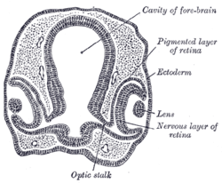
Transverse section of head of chick embryo of fifty-two hours’ incubation.
Gray865.png
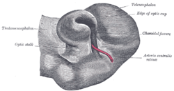
Optic cup and choroidal fissure seen from below, from a human embryo of about four weeks. (Optic stalk labeled at center left.)
Latin pedunculus opticus
Gray's subject #224 1001
Carnegie stage 14
Code TE E5.14.3.4.2.2.6

The optic vesicles project toward the sides of the head, and the peripheral part of each expands to form a hollow bulb, while the proximal part remains narrow and constitutes the optic stalk.

Closure of the choroid fissure in the optic stalk occurs during the seventh week of development. The former optic stalk is then called the optic nerve.[1] The Bottom Line: the optic stalks are the structures that precede the optic nerves embryologically.

References

  1. ^ Kaplan Qbook - USMLE Step 1 - 5th edition - page 55

External links

This article was originally based on an entry from a public domain edition of Gray's Anatomy. As such, some of the information contained within it may be outdated.


Wikimedia Foundation. 2010.

Игры ⚽ Поможем написать реферат

Look at other dictionaries:

  • optic stalk — n the constricted part of the optic vesicle by which it remains continuous with the embryonic forebrain * * * a slender structure attaching the optic vesicle, and later the optic cup, to the brain wall in the embryo …   Medical dictionary

  • optic stalk — noun : the constricted part of the optic vesicle by which it remains continuous with the embryonic forebrain …   Useful english dictionary

  • Optic vesicle — Transverse section of head of chick embryo of forty eight hours’ incubation. (Optic vesicle labeled at lower right.) …   Wikipedia

  • Optic cup (embryology) — Transverse section of head of chick embryo of forty eight hours’ incubation. (Margin of optic cup labeled at upper right.) …   Wikipedia

  • Optic vesicles — Infobox Embryology Name = PAGENAME Latin = v. ophthalmica GraySubject = 224 GrayPage = 1001 Caption = Transverse section of head of chick embryo of forty eight hours’ incubation. (Optic vesicle labeled at lower right.) Caption2 = Human embryo… …   Wikipedia

  • stalk — A narrowed connection with a structure or organ. allantoic s. the narrow connection between the intraembryonic portion of the allantois and the extraembryonic allantoic vesicle …   Medical dictionary

  • Optic cup — The white, cup like area in the center of the optic disc. The optic disk is the circular area at the back of the inside of the eye where the optic nerve connects to the retina. * * * optic cup n the optic vesicle after invaginating to form a 2… …   Medical dictionary

  • coloboma of optic disk — coloboma of optic nerve 1. a coloboma, mild or severe, within or at the optic nerve head. A mild coloboma may be a separate, isolated entity, unilateral, and limited to minor cupping in the optic disk. A severe coloboma may be part of a bridge… …   Medical dictionary

  • human embryology — ▪ biology Introduction       the process encompassing the period from the formation of an embryo, through the development of a fetus, to birth.       The human body, like that of most animals, develops from a single cell produced by the union of… …   Universalium

  • Embryology — 1 morula, 2 blastula 1 blastula, 2 gastrula with blastopor …   Wikipedia

Share the article and excerpts

Direct link
Do a right-click on the link above
and select “Copy Link”