- Lateral horn
Infobox Anatomy
Name = Lateral horn
Latin = cornu laterale medullae spinalis
GraySubject = 185
GrayPage = 753
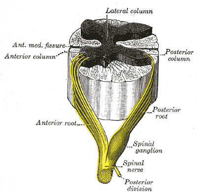
Caption = A spinal nerve with its anterior and posterior roots. (Lateral column labeled at top.)
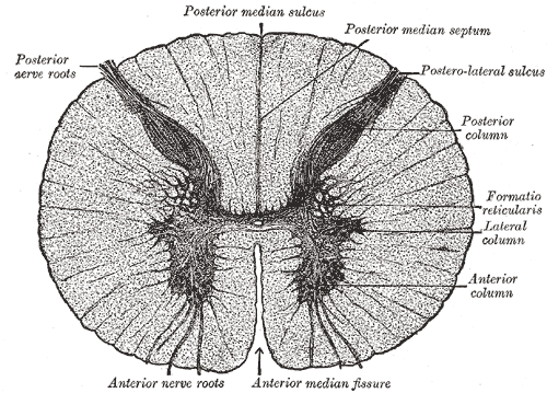
Caption2 = Transverse section of the medulla spinalis in the mid-thoracic region. (Lateral column labeled at center right.)
System =
Precursor =
MeshName =
MeshNumber =
DorlandsPre = c_55
DorlandsSuf = 12259818
In thethoracic region, the postero-lateral part of theanterior column projects lateralward as a triangular field, which is named the lateral column (lateral cornu, lateral horn).Nerve Cells in the Lateral Column
These form a column (the intermedioloateral cell column) which is best marked where the lateral gray column is differentiated, viz., in the thoracic region; but it can be traced throughout the entire length of the
medulla spinalis in the form of groups of small cells which are situated in the anterior part of theformatio reticularis . Theintermediolateral cell column exists at vertebral levels T1 - L2 and mediates the entire sympathetic innervation of the body. Preganglionic, myelinated GVA fibers from viscera course through prevertebral and paravertebral (sympathetic) ganglia, white rami and dorsal roots to synapse with cells of the intermediolateral cell column. These cells then give rise to preganglionic GVE fibers which will pass through ventral spinal roots, white rami, and into paravertebral ganglia where some will synapse, thus sending unmyelinated, postganglionic fibers through gray rami and into peripheral nerves. Those fibers that do not synapse in the paravertebral ganglia will eventually synapse at prevertebral ganglia near target viscera. Postganglionic neurons in the prevertebral ganglia send postganglionic fibers to target tissues.In the upper part of the cervical region and lower part of the
medulla oblongata as well as in the third and fourth sacral segments this column is again differentiated.In the medulla it is known as the
lateral nucleus .The cells of this column are fusiform or star-shaped, and of a medium size: the axons of some of them pass into the
anterior nerve roots , by which they are carried to thesympathetic nerves : they constitute thewhite rami and aresympathetic orvisceral efferent fibers ; they are also known aspreganglionic fibers of the sympathetic system ; the axons of others pass into the anterior andlateral funiculi , where they become longitudinal.
Wikimedia Foundation. 2010.