Medial epicondyle of the femur

Medial epicondyle of the femur
Bone: Medial epicondyle of the femur
Gray345.png
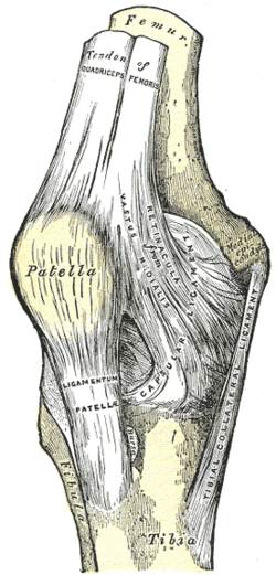
Right knee-joint. Anterior view. (Medial epicondyle visible at right.)
Gray244.png
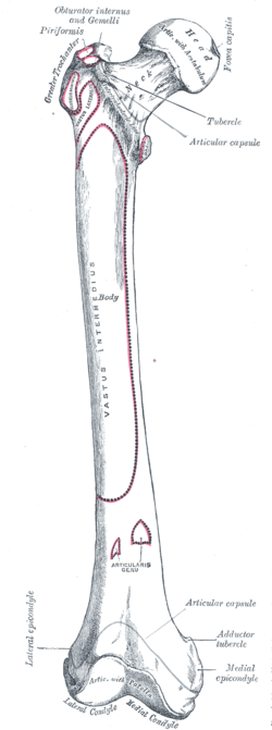
Right femur. Anterior surface. (Medial epicondyle labeled at bottom right.)
Latin epicondylus medialis femoris
Gray's subject #59 247

The medial epicondyle of the femur is a bony protrusion located on the medial side of the bone's distal end.

Located above the medial condyle, it bears an elevation, the adductor tubercle,[1] which serves for the attachment of the superficial part, or "tendinous insertion", of the adductor magnus[2]. This tendinous part here forms an intermuscular septum which forms the medial separation between the thigh's flexors and extensors.[3]

Behind it, and proximal to the medial condyle[4] is a rough impression which gives origin to the medial head of the Gastrocnemius.

Contents

See also

Notes

  1. ^ Platzer (2004), p 192
  2. ^ Thieme Atlas of Anatomy (2006), p 426
  3. ^ Platzer (2004), 9 206
  4. ^ Platzer (2004), p 262

References

  • Platzer, Werner (2004). Color Atlas of Human Anatomy, Vol. 1: Locomotor System (5th ed.). Thieme. ISBN 3-13-533305-1. 
  • Thieme Atlas of Anatomy: General Anatomy and Musculoskeletal System. Thieme. 2006. ISBN 1-58890-419-9. 

External links

This article was originally based on an entry from a public domain edition of Gray's Anatomy. As such, some of the information contained within it may be outdated.


Wikimedia Foundation. 2010.

Игры ⚽ Нужна курсовая?

Look at other dictionaries:

  • Lateral epicondyle of the femur — Infobox Bone Name = Lateral epicondyle of the femur Latin = e. lateralis femoris GraySubject = 59 GrayPage = 247 Caption = Lower extremity of right femur viewed from below. (Lateral epicondyle visible at left.) Caption2 = Right femur. Anterior… …   Wikipedia

  • Medial epicondyle — can refer to: Medial epicondyle of the humerus (ventral epicondyle in birds) Medial epicondyle of the femur This disambiguation page lists articles associated with the same title. If an …   Wikipedia

  • medial collateral ligament — n 1) a ligament that connects the medial epicondyle of the femur with the medial condyle and medial surface of the tibia and that helps to stabilize the knee by preventing lateral dislocation called also MCL, tibial collateral ligament compare… …   Medical dictionary

  • Medial collateral ligament — Ligament: Medial collateral ligament Diagram of the right knee. (Medial collateral ligament labeled at center right.) Latin ligamentum collaterale tibiale …   Wikipedia

  • epicondyle — A projection from a long bone near the articular extremity above or upon the condyle. SYN: epicondylus [TA]. [epi + G. kondylos, a knuckle] lateral e. of femur [TA] the epicondylus located proximal to the …   Medical dictionary

  • Muscular system of the horse — See also: Equine anatomy Contents 1 Types of muscle 2 Build of skeletal muscle 3 Tendons of the lower leg 4 Main skeletal muscles of the horse …   Wikipedia

  • Medial condyle of femur — Bone: Medial condyle of femur Lower extremity of right femur viewed from below …   Wikipedia

  • List of muscles of the human body — Skeletal muscles homo sapiens Muscles of the human body: Overview Head  |  Neck  |&# …   Wikipedia

  • Lower extremity of femur — Infobox Bone Name = Lower extremity of femur Latin = GraySubject = 59 GrayPage = 243 Caption = Lower extremity of right femur viewed from below. Caption2 = Left knee joint from behind, showing interior ligaments. System = MeshName = MeshNumber =… …   Wikipedia

  • Ligament — A ligament is a tough band of connective tissue that connects various structures such as two bones. Ligament is a fitting term; it comes from the Latin ligare meaning to bind or tie. * * * 1. A band or sheet of fibrous tissue connecting two or… …   Medical dictionary

Share the article and excerpts

Direct link
Do a right-click on the link above
and select “Copy Link”