- Basilar membrane
Infobox Anatomy
Name = PAGENAME
Latin = lamina basilaris ductus cochlearis
GraySubject = 232
GrayPage = 1056
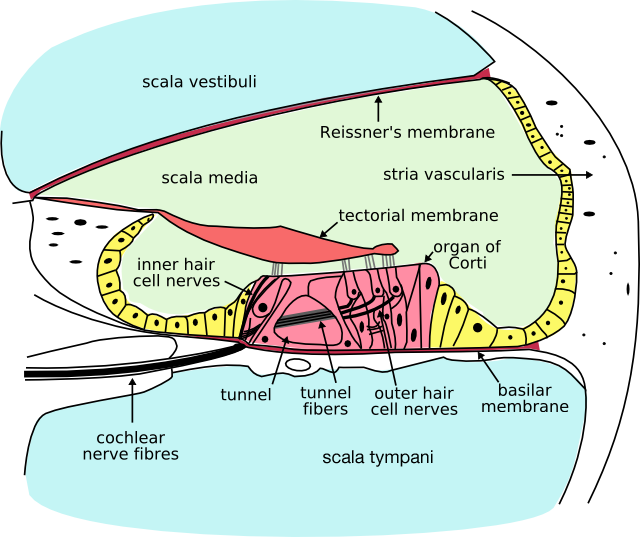
Caption = Cross section of the cochlea.
Caption2 =
Precursor =
System =
Artery =
Vein =
Nerve =
Lymph =
MeshName = Basilar+membrane
MeshNumber = A09.246.631.246.125
DorlandsPre = l_02
DorlandsSuf = 12475936
The basilar membrane within thecochlea of theinner ear is a stiff structural element that separates two liquid-filled tubes that run along the coil of the cochlea, thescala media and thescala tympani (see figure).Function
Endolymph/perilymph separation
The fluids in these two tubes, the
endolymph and theperilymph are very different chemically, biochemically, and electrically. Therefore they are kept strictly separated. This separation is the main function ofReissner's membrane (betweenscala vestibuli and scala media), and is one of the functions of the basilar membrane in the hearing organ of all landvertebrates .A "base" for the sensory cells
The basilar membrane is also the "base" for the sensory cells of hearing, the
hair cells (see figure). This function gave the "basilar" membrane its name, and it is again present in all land vertebrates. Due to its location, the basilar membrane places the hair cells in a position where they are adjacent to both the endolymph and the perilymph, which is a precondition of hair cell function.Frequency dispersion
A third, evolutionarily younger, function of the basilar membrane is strongly developed in the cochlea of most mammalian species and weakly developed in some bird species. It is the function of frequency
dispersion of incoming sound waves. In brief, the membrane is tapered and it is stiffer at one end than at the other. The dispersion of fluid waves causes sound input of a certain frequency to vibrate some locations of the membrane more than other locations. As shown in experiments by Nobel Prize laureateGeorg von Békésy , high frequencies lead to maximum vibrations at the basal end of the cochlear coil (narrow, stiff membrane), and low frequencies lead to maximum vibrations at the apical end of the cochlear coil (wide, more compliant membrane). This "place-frequency map" can be described quantitatively by theGreenwood Function and its variants.Anatomy
The basilar membrane is a pseudo-resonant structure [M. Holmes and J. D. Cole, "Pseudoresonance in the cochlea, ' in: "Mechanics of Hearing", E. de Boer and M. A. Viergever (editors), Proceedings of the IUTAM/ICA Symposium, Delft (1983), pp. 45-52.] that, like strings on an instrument, varies in width and stiffness. The "string" of the basilar membrane is not a set of parallel strings, as in a guitar, but a long structure that has different properties (width, stiffness, mass, damping, and the dimensions of the ducts that it couples to) at different points along its length. The motion of the basilar membrane is generally described as a traveling wave. [cite book | title = Compression: From Cochlea to Cochlear Implants | author = Richard R. Fay, Arthur N. Popper, and Sid P. Bacon | publisher = Springer | year = 2004 | isbn = 0387004963 | url = http://books.google.com/books?id=4Z5mV_qhB5kC&pg=PA4&ots=S8aTl5rdCm&dq=traveling-wave++%22basilar+membrane%22&as_brr=3&sig=hSmHnapwyixPRqbm_HtYJohBycU#PPA4,M1 ] The parameters of the membrane at a given point along its length determine its characteristic frequency (CF), the frequency at which it is most sensitive to sound vibrations. The Basilar membrane is widest (0.42–0.65 mm) and least taut at the apex of the cochlea, and narrowest (0.08–0.16 mm) and most taut at the base. [Oghalai JS. The cochlear amplifier: augmentation of the traveling wave within the inner ear. "Current Opinion in Otolaryngology & Head & Neck Surgery". 12(5):431-8, 2004] High-frequency sounds localize near the base of the cochlea (near the round and oval windows), while low-frequency sounds localize near the apex.
=AdditionalReferences
*Fritzsch B: The water-to-land transition: Evolution of the tetrapod basilar papilla; middle ear, and auditory nuclei. In: cite book |author=Douglas B. Webster, Richard R. Fay, Arthur N. Popper, editors |title=The Evolutionary biology of hearing |publisher=Springer-Verlag |location=Berlin |year=1992 |pages=351-375 |isbn=0-387-97588-8 |oclc= |doi=
*
*
*
*
External links
*
* [http://www.iurc.montp.inserm.fr/cric/audition/english/cochlea/fcochlea.htm Functional anatomy of the inner ear: plenty of images, animations, and very concise functional explanations]
Wikimedia Foundation. 2010.