- Thoracic wall
-
Thoracic wall 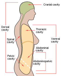
Body cavities 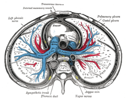
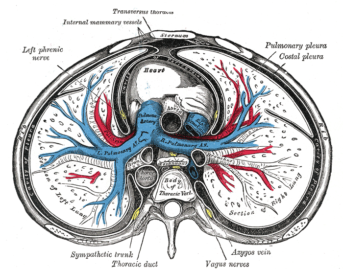
A transverse section of the thorax, showing the contents of the middle and the posterior mediastinum. The thoracic wall (or chest wall) is the boundary of the thoracic cavity.
The bony portion is known as the thoracic cage. However, the wall also includes muscle, skin, and fascia.
External links
- MeSH Thoracic+wall
- SUNY Labs 18:lo-0000
- chest+wall at eMedicine Dictionary
- Anatomy stubs
- Thorax

Categories:
Wikimedia Foundation. 2010.