- Corticospinal tract
-
"Pyramidal tract" redirects here. This page refers to the nerve fibres underlying the pyramids. For the actual area of the brain, Pyramids, see Pyramid of medulla oblongata.
Brain: Corticospinal tract 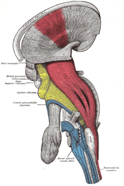
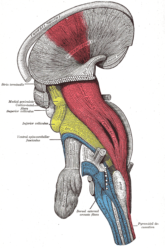
Deep dissection of brain-stem. Lateral view. ("pyramidal tract" visible in red, and "pyramidal decussation" labeled at lower right.) 
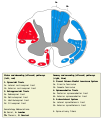
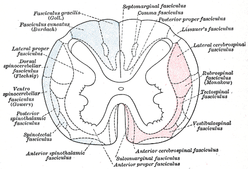
Diagram of the principal fasciculi of the spinal cord. Latin tractus corticospinalis Gray's subject #185 759 NeuroNames ancil-373 MeSH Pyramidal+Tracts NeuroLex ID birnlex_1464 The corticospinal or pyramidal tract is a collection of axons that travel between the cerebral cortex of the brain and the spinal cord.
The corticospinal tract mostly contains motor axons. It actually consists of two separate tracts in the spinal cord: the lateral corticospinal tract and the anterior corticospinal tract. An understanding of these tracts leads to an understanding of why for the most part, one side of the body is controlled by the opposite side of the brain.
The corticobulbar tract is also considered to be a pyramidal tract, though it carries signals to motor neurons of the cranial nerve nuclei, rather than the spinal cord.[1]
The neurons of the corticospinal tracts are sometimes referred to as pyramidal tract neurons (PTN), because their axons form part of the pyramidal tracts leading to the spinal cord, which in turn are named such because in cross-section they resemble pyramids as they pass through the medulla. [2]
Pyramidal tract neurons, however, are not to be confused with pyramidal neurons: a super-class of neurons, found in many parts of the brain including the cerebral cortex and the hippocampus, whose name derives from the pyramid-like shape of the cell body (soma). In fact, corticospinal neurons are both pyramidal tract neurons (because their axons pass through the medullary pyramids) and pyramidal neurons (because their cell body is shaped like a pyramid).
The corticospinal tract is concerned specifically with discrete voluntary skilled movements, especially of the distal parts of the limbs (sometimes called "fractionated" movements).
Contents
The motor pathway
The corticospinal tract originates from pyramidal cells in layer V of the cerebral cortex. About half of its fibres arise from the primary motor cortex. Other contributions come from the supplementary motor area, premotor cortex, somatosensory cortex, parietal lobe, and cingulate gyrus. The average fiber diameter is in the region of 10μm; around 3% of fibres are extra-large (20μm) and arise from Betz cells, mostly in the leg area of the primary motor cortex.
Upper motor neurons
The neuronal cell bodies in the motor cortex, together with their axons that travel down through the brain stem and spinal cord are commonly referred to as upper motor neurons. It should be noted however, that they do not project to muscles, and thus the term 'motor neuron' is somewhat misleading.
Decussation and synapses
Some of the neuronal cell bodies in the motor cortex send long axons to the motor cranial nerve nuclei mainly of the contralateral side of the midbrain (cortico-mesencephalic tract), pons (Corticopontine tract), and medulla oblongata (cortico-bulbar tract), decussating just before they reach their target nuclei. These are called geniculate fibers. Many more motor cortex neurons, however, extend fibers all the way down to the spinal cord (corticospinal tract).
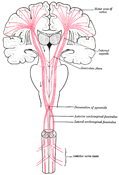
- Most of the corticospinal fibers (about 80%) cross over to the contralateral side in the medulla oblongata (pyramidal decussation). Those that cross in the medulla oblongata travel in the lateral corticospinal tract.
- 10% enter the lateral corticospinal tract on the same side.
- The remainder of them (10%) cross over at the level that they exit the spinal cord, and these travel in the anterior corticospinal tract.
Whichever of these two tracts it travels in, a corticospinal axon will synapse with another neuron in the ventral horn. This ventral horn neuron is considered a second-order neuron in this pathway, but is not part of the corticospinal tract itself.
From cerebral to motor neurons
The motor axons move closer together as they travel down through the cerebral white matter, and form part of the posterior limb of the internal capsule.
The motor fibers continue down into the brainstem. The bundle of corticospinal axons is visible as two column-like structures ("pyramids") on the ventral surface of medulla oblongata - this is where the name pyramidal tract comes from.
After the decussation, the axons travel down the spinal cord as the lateral corticospinal tract. Fibers that do not cross over in the medulla oblongata travel down the separate anterior corticospinal tract, and most of them cross over to the contralateral side in the spinal cord, shortly before reaching the lower motor neurons.
 Horizontal section through the lower part of the pons, showing the fibers of the corticospinal tract (#19) passing through the pontine nuclei
Lower motor neurons
In the spinal cord, the axons of the upper motor neuron connect (most of them via interneurons, but to a lesser extent also via direct synapses) with the lower motor neurons, located in the ventral horn of the spinal cord.
In the brain stem, the lower motor neurons are located in the motor cranial nerve nuclei (oculomotor, trochlear, motor nucleus of the trigeminal nerve, abducens, facial, accessory, hypoglossal). The lower motor neuron axons leave the brain stem via motor cranial nerves and the spinal cord via anterior roots of the spinal nerves respectively, end-up at the neuromuscular plate and provide motor innervation for voluntary muscles.
Sensory pathways
- Spinothalamic tract
- Spinocerebellar tract
- Visual pathway
- Olfactory system
- Posterior column pathway
Corticospinal tract damage
Damage to the descending motor pathways anywhere along the trajectory from the cerebral cortex to the lower end of the spinal cord gives rise to a set of symptoms called the "Upper Motor Neuron Syndrome". A few days after the injury to the upper motor neurons a pattern of motor signs and symptoms appears, including spasticity, the decreased vigor (and increased threshold) of superficial reflexes, a loss of the ability to perform fine movements, and an extensor plantar response known as the Babinski sign.[3]
Extrapyramidal motor pathways
These are motor pathways that lie outside the corticospinal tract and are beyond voluntary control. Their main function is to support voluntary movement and help control posture and muscle tone. See extrapyramidal motor system.
Additional images
-
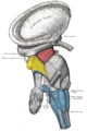
Dissection of brain-stem. Lateral view.
-

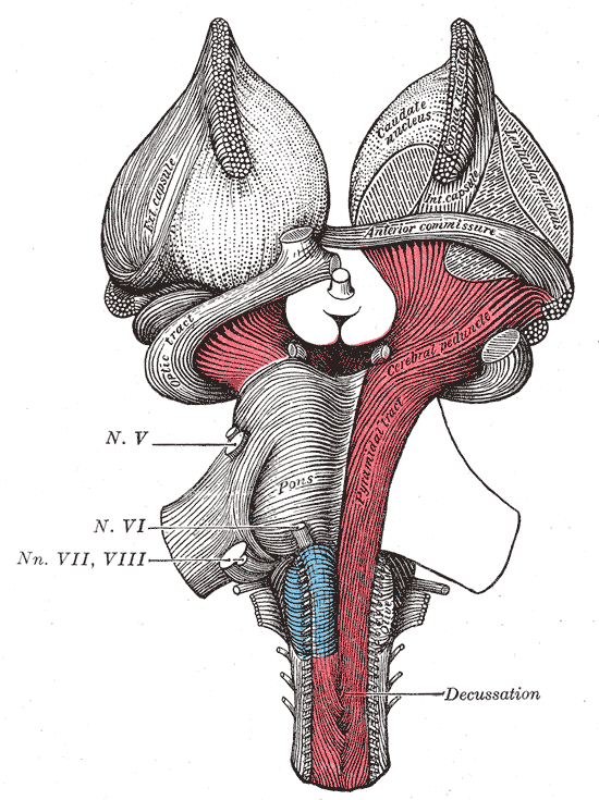
Superficial dissection of brain-stem. Ventral view.
-
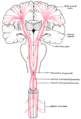
The motor tract.
-
-

Spinal cord tracts
References
- ^ Chapter 9 of "Principles of Physiology" (3rd edition) by Robert M. Berne and Mathew N. Levy. Published by Mosby, Inc. (2000) ISBN:0-323-00813-5.
- ^ The Brain From Top To Bottom
- ^ http://www.ncbi.nlm.nih.gov/bookshelf/br.fcgi?book=neurosci&part=A1191
External links
- 959774759 at GPnotebook
- Brainstem at UWisc 01Pyramid
- McGill
- BrainMaps at UCDavis Corticospinal tract
Human brain: mesencephalon (midbrain) (TA A14.1.06, GA 9.800) Tectum
(Dorsal)SurfaceWhite: Sensory/ascendingWhite: Motor/descendingPeduncle
(Ventral)White: Sensory/ascendinglemnisci (Medial, Lateral) · Ascending MLF (Vestibulo-oculomotor fibers) · Spinothalamic tract · Anterior trigeminothalamic tract · Dentatothalamic tractWhite: Motor/descendingGrey: otherPeriaqueductal gray/Raphe nuclei (Dorsal raphe nucleus)
Ventral tegmental area • Pedunculopontine nucleus • Red nucleus
riMLFBaseWhite: Motor/descendingCerebral crus: Corticospinal tract · Corticobulbar tract · Corticopontine tract/Frontopontine fibers/Temporopontine fibersSurfaceHuman brain, rhombencephalon, metencephalon: pons (TA A14.1.05.101–604, GA 9.785) Dorsal/
(tegmentum)SurfaceWhite: Sensory/ascendingTrapezoid body/VIII · Trigeminal lemniscus (Dorsal trigeminal tract, Ventral trigeminal tract) · Medial lemniscus · Lateral lemniscus
MLF, III, IV and VI: Vestibulo-oculomotor fibers
Anterior trigeminothalamic tract · Central tegmental tractWhite: Motor/descendingICP (Vestibulocerebellar tract)
MLF, III, IV and VI: Vestibulospinal tract (Medial vestibulospinal tract, Lateral vestibulospinal tract)Other greyVentral/
(base)White: Motor/descendingSurfaceBasilar sulcusOther grey: Raphe/
reticularAnatomy of torso (primarily): the spinal cord (TA 14.1.02, GA 9.749) External, dorsal Posterior median sulcus · Posterolateral sulcusGrey matter/
Rexed laminaeI–VI: Posterior hornI: Marginal nucleus · II: Substantia gelatinosa of Rolando · III+IV: Nucleus proprius · Spinal lamina V · Spinal lamina VIVII: Lateral hornVIII–IX: Anterior hornX: OtherWhite matter somatic/
ascending
(blue)Posterior/PCML: touch: Gracile · Cuneate
Lateral: proprioception: Spinocerebellar (Dorsal, Ventral) · pain/temp: Spinothalamic (Lateral, Anterior) · Posterolateral (Lissauer) · Spinotectal
Spinoreticular tract · Spino-olivary tractmotor/
descending
(red)Lateral: Corticospinal (Lateral) · Ep (Rubrospinal, Olivospinal)
Anterior: Corticospinal (Anterior) · Ep (Vestibulospinal, Reticulospinal, Tectospinal)bothExternal, ventral Anterior median fissure · Anterolateral sulcusExternal, general Categories:- Central nervous system pathways
- Motor system


Wikimedia Foundation. 2010.