- Paraventricular nucleus of hypothalamus
Infobox Brain
Name = PAGENAME
Latin = nuclei paraventricularis hypothalami
GraySubject =
GrayPage =

Caption = Human paraventricular nucleus (PVN) in this coronal section is indicated by the shaded area. Dots representvasopressin (AVP) neurons (also seen in thesupraoptic nucleus , SON). The medial surface is the 3rd ventricle (3V).
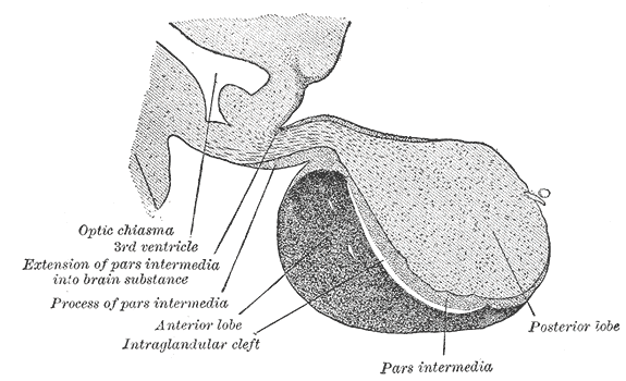
Caption2 = Magnocellular neurons of the PVN and SON project to the posterior "lobe" of the pituitary
IsPartOf =
Components =
Artery =
Vein =
Acronym =
BrainInfoType = hier
BrainInfoNumber = 370
MeshName = Paraventricular+hypothalamic+nucleus
MeshNumber = A08.186.211.730.385.357.342.400
DorlandsPre = n_11
DorlandsSuf = 12582324
The paraventricular nucleus (PVN) is an aggregation of neurons in thehypothalamus which produces manyhormones .Location
The paraventricular nucleus lies adjacent to the
third ventricle , from which it derives its name, "paraventricular" meaning "alongside a ventricle."It does lie within the periventricular zone and must not be confused with the
periventricular nucleus , which occupies a more medial position, beneath thethird ventricle .The PVN is highly vascularised and is protected by the
blood-brain barrier , although its neuroendocrine neurons extend to sites (in themedian eminence and in theposterior pituitary ) beyond the blood-brain barrier.Neurons
The PVN contains
magnocellular neurosecretory cell s whose axons extend into theposterior pituitary , parvocellular neurosecretory cells that project to themedian eminence , and several populations ofpeptide -containing cells that project to many different brain regions.Magnocellular neurosecretory neurons
The
magnocellular cells in the PVN elaborate and secrete two peptidehormones ,oxytocin andvasopressin .These hormones are packaged into large vesicles, which are then transported down the
axon s of the cells and released from neurosecretory nerve terminals residing in theposterior pituitary gland .Similar magnocellular neurons are found in the
supraoptic nucleus .Parvocellular neurosecretory neurons
The axons of the
parvocellular neurosecretory neurons of the PVN project to the median eminence, at the base of the brain, where their neurosecretory nerve terminals release peptides into blood vessels in thehypothalamo-pituitary portal system . The blood vessels carry the peptides to theanterior pituitary gland, where they regulate the secretion of hormones into the systemic circulation. The parvocellular neurosecretory cells include those which make*
Corticotropin-releasing hormone (CRH), which regulatesACTH secretion from theanterior pituitary gland ,
*Vasopressin , which also regulates ACTH secretion (vasopressin and CRH act synergistically to stimulate ACTH secretion), and
*Thyrotropin-releasing hormone (TRH), which regulatesTSH andprolactin secretion.Centrally-projecting neurons
As well as neuroendocrine neurons, the PVN contains
interneuron s and populations of neurons that project centrally (i.e., to other brain regions). The centrally-projecting neurons include* Parvocellular oxytocin cells, which project mainly to the
brainstem andspinal cord and are involved, respectively, in gastric reflexes and penile erection,
* Parvocellular vasopressin cells, which project to many points in the hypothalamus andlimbic system , as well as to the brainstem and spinal cord (these are involved in blood pressure and temperature regulation), and
* Parvocellular CRH neurons, which are thought to be involved in stress-related behaviors.Afferent inputs to the PVN
The PVN receives afferent inputs from many brain regions.
Among these, inputs from neurons in structures adjacent to the anterior wall of the third ventricle (the "AV3V region") carry information about the electrolyte composition of the blood, and about circulating concentrations of such hormones as
angiotensin andrelaxin , to regulate the magnocellular neurons.Inputs from the brainstem (the
nucleus of the solitary tract ) and the ventrolateral medulla carry information from the heart andstomach . Inputs from thehippocampus to the CRH neurones are important regulators of stress responses.Inputs from
neuropeptide Y -containing neurons in thearcuate nucleus co-ordinate metabolic regulation (via TRH secretion) with regulation of energy intake.References
External links
* [http://www.painresearch.utah.edu/cancerpain/ch06f2.html Diagram of location in brain at painresearch.utah.edu]
Wikimedia Foundation. 2010.