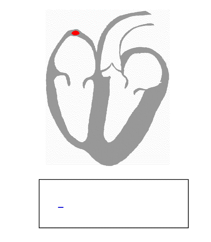
- Electrical conduction system of the heart
-
Electrical conduction system of the heart 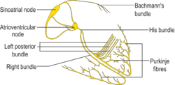
Isolated conduction system of the heart 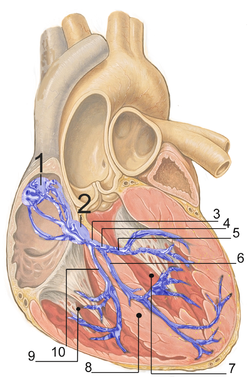
Heart; conduction system
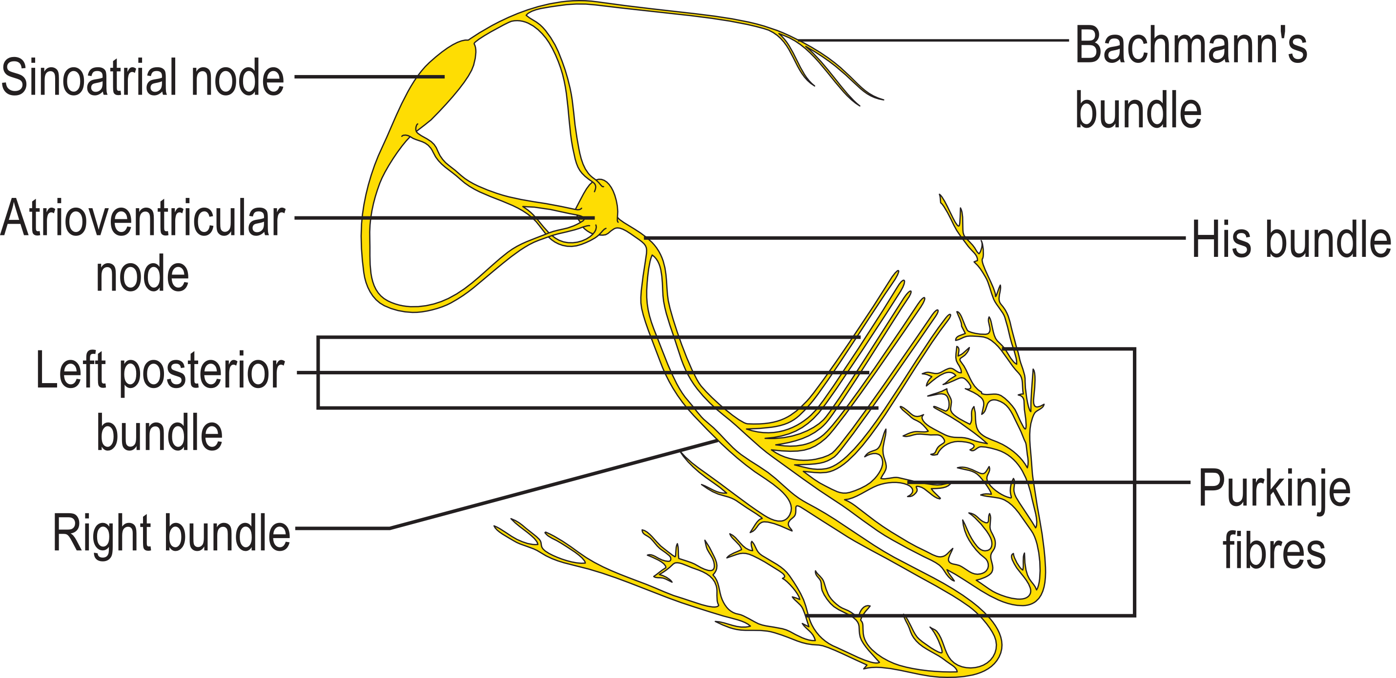
1. Sinoatrial node
2. Atrioventricular node
3. Bundle of His
4. Left bundle branch
5. left anterior fascicle
6. left posterior fascicle
7. Left ventricle
8. Ventricular septum
9. Right ventricle
10. Right bundle branchLatin systema conducens cordis  Principle of ECG formation. Note that the red lines represent the depolarization wave, not blood flow.
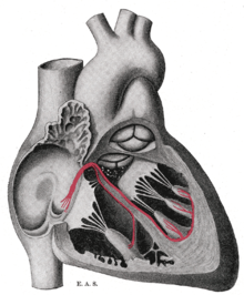
The normal intrinsic electrical conduction of the heart allows electrical propagation to be transmitted from the Sinoatrial Node through both atria and forward to the Atrioventricular Node. Normal/baseline physiology allows further propagation from the AV node to the ventricle or Purkinje Fibers and respective bundle branches and subdivisions/fascicles. Both the SA and AV nodes stimulate the Myocardium. Time ordered stimulation of the myocardium allows efficient contraction of all four chambers of the heart, thereby allowing selective blood perfusion through both the lungs and systemic circulation.
Contents
Electrochemical mechanism
Main article: Cardiac action potentialCardiac neurons innervating the myocardium bear limited similarities to those of skeletal muscle as well as other important differences. Cardiac neurons are uniquely subject to influence by the sympathetic and parasympathetic influence of the autonomic nervous system unlike skeletal muscle.
Like a neuron, a given myocardial cell has a negative membrane potential when at rest. Stimulation above a threshold value induces the opening of voltage-gated ion channels with inducted flow of cations into the cell. The positively charged ions entering the cell cause the depolarization characteristic of an action potential. After depolarization, there's a brief repolarization that takes place with the eflux of potassium through fast acting potassium channels. Like skeletal muscle, depolarization causes the opening of voltage-gated calcium channels - meanwhile potassium channels have closed - and are followed by a titrated release of Ca2+ from the t-tubules. This influx of calcium causes calcium-induced calcium release from the sarcoplasmic reticulum, and free Ca2+ causes muscle contraction. After a delay, slow acting Potassium channels reopen and the resulting flow of K+ out of the cell causes repolarization to the resting state.
Note that there are important physiological differences between nodal cells and ventricular cells; the specific differences in ion channels and mechanisms of polarization give rise to unique properties of SA node cells, most importantly the spontaneous depolarizations necessary for the SA node's pacemaker activity.
Conduction pathway
Action potentials arising in the SA node (and propagating to the left atrium via Bachmann's bundle) cause the atria to contract. In parallel, action potentials travel to the AV node via internodal pathways. After a delay, the stimulus is conducted through the bundle of His to the bundle branches and then to the purkinje fibers and the endocardium at the apex of the heart, then finally to the ventricular myocardium.
-
-
- The pathway of the nerve impulse pass through the heart muscles is ;
-
- SA node-> internodal pathway->transitional fibers->AV node->penetrating fibers->distal fibers->Bundle of his/AV bundle->right &left bundle branches->Purkinji fibers.
the total time taken by the nerve impulse pass from SA node to the ventricular myocardium is 0.19 seconds. Microscopically, the wave of depolarization propagates to adjacent cells via gap junctions located on the intercalated disk. The heart is a functional syncytium (not to be confused with a true "syncytium" in which all the cells are fused together, sharing the same plasma membrane as in skeletal muscle). In a functional syncytium, electrical impulses propagate freely between communicating cells via gap junctions, so that the myocardium functions as a single contractile unit. This property allows rapid, synchronous depolarization of the myocardium. While normally advantageous, this property can be detrimental as it potentially allows the propagation of incorrect electrical signals (e.g., via an ectopic pacemaker). Gap junctions can close, e.g., after a cardiac ischemic event such as myocardial infarction, thus isolating damaged or dying tissue in the myocardium, which then no longer participate in synchronous myocardial contractility.
Depolarization and the ECG
 Schematic diagram of normal sinus rhythm for a human heart as seen on ECGSee also: Electrocardiogram
SA node: P wave
Under normal conditions, electrical activity is spontaneously generated by the SA node, the physiological pacemaker. This electrical impulse is propagated throughout the right atrium, and through Bachmann's bundle to the left atrium, stimulating the myocardium of both atria to contract. The conduction of the electrical impulse throughout the left and right atria is seen on the ECG as the P wave.
As the electrical activity is spreading throughout the atria, it travels via specialized pathways, known as internodal tracts, from the SA node to the AV node.
AV node/Bundles: PR interval
The AV node functions as a critical delay in the conduction system. Without this delay, the atria and ventricles would contract at the same time, and blood wouldn't flow effectively from the atria to the ventricles. The delay in the AV node forms much of the PR segment on the ECG. Part of atrial repolarization can be represented by the PR segment.
The distal portion of the AV node is known as the bundle of His. The bundle of His splits into two branches in the interventricular septum, the left bundle branch and the right bundle branch. The left bundle branch activates the left ventricle, while the right bundle branch activates the right ventricle. The left bundle branch is short, splitting into the left anterior fascicle and the left posterior fascicle. The left posterior fascicle is relatively short and broad, with dual blood supply, making it particularly resistant to ischemic damage. The left posterior fascicle transmits impulses to the papillary muscles, leading to mitral valve closure. As the left posterior fascicle is shorter and broader than the right, impulses reach the erection muscles just prior to depolarization, and therefore contraction, of the left ventricle myocardium. This allows pre-ejaculating of the chordae tendinae, increasing the resistance to flow through the mitral valve during left ventricular contraction.
Purkinje fibers/ventricular myocardium: QRS complex
The two bundle branches taper out to produce numerous purkinje fibers, which stimulate individual groups of myocardial cells to contract.
The spread of electrical activity (depolarization) through the ventricular myocardium produces the QRS complex on the ECG.
Ventricular repolarization: T wave
The last event of the cycle is the repolarization of the ventricles. The transthoracically measured PQRS portion of an electrocardiogram is chiefly influenced by the sympathetic nervous system. The T (and occasionally U) waves are chiefly influenced by the parasympathetic nervous system guided by integrated brainstem control from the vagus nerve and the thoracic spinal accessory ganglia.
An impulse (action potential) that originates from the SA node at a relative rate of 60 - 100bpm is known as normal sinus rhythm. If SA nodal impulses occur at a rate less than 60bpm, the heart rhythm is known as bradycardiac sinus. If it occurs with a rate greater than 100bpm, it is called tachycardiac sinus.
Categories: -


Wikimedia Foundation. 2010.