Interosseous membrane of the leg
- Interosseous membrane of the leg
Infobox Anatomy
Name = PAGENAME
Latin = membrana interossea cruris, ligamentum tibiofibulare medium
GraySubject = 94
GrayPage = 348
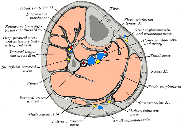
Caption = Cross-section through middle of left leg. (Interosseus membrane labeled at upper right.)
Caption2 =
System =
MeshName =
MeshNumber =
DorlandsPre = m_08
DorlandsSuf = 12522106
The interosseous membrane of the leg (middle tibiofibular ligament) extends between the interosseous crests of the tibia and fibula, and separates the muscles on the front from those on the back of the leg.
It consists of a thin, aponeurotic lamina composed of oblique fibers, which for the most part run downward and lateralward; some few fibers, however, pass in the opposite direction.
It is broader above than below. Its upper margin does not quite reach the tibiofibular joint, but presents a free concave border, above which is a large, oval aperture for the passage of the anterior tibial vessels to the front of the leg.
In its lower part is an opening for the passage of the anterior peroneal vessels.
It is continuous below with the interosseous ligament of the tibiofibular syndesmosis, and presents numerous perforations for the passage of small vessels.
It is in relation, in front, with the Tibialis anterior, Extensor digitorum longus, Extensor hallucis proprius, Peronæus tertius, and the anterior tibial vessels and deep peroneal nerve; behind, with the Tibialis posterior and Flexor hallucis longus.
=Additional
External links
*
* [http://www.massagetherapy.com/articles/index.php/article_id/496 Diagrams at massagetherapy.com]
Wikimedia Foundation.
2010.
Look at other dictionaries:
Interosseous membrane of leg — Cross section through middle of left leg. (Interosseus membrane labeled at upper right.) Latin membrana interossea cruris, ligamentum tibiofibulare medium Gray s … Wikipedia
Interosseous membrane — An interosseous membrane is a broad and thin plane of fibrous tissue that separates many of the bones of the body. It is an important component of many joints.Interosseous membranes in the human body: * Interosseous membrane of the forearm *… … Wikipedia
Fascial compartments of leg — Cross section through middle of left leg. (Colours correspond to fascial compartments; red text names muscles in each compartment; grey text names neurovascular structures in each compartment) On the human body, the limbs can be divided into… … Wikipedia
membrane — 1. A thin sheet or layer of pliable tissue, serving as a covering or envelope of a part, as the lining of a cavity, as a partition or septum, or to connect two structures. SYN: membrana [TA]. 2. SYN: biomembrane. [L. membrana, a skin or m … Medical dictionary
Skeletal system of the horse — The skeletal system has three major functions in the body. It protects vital organs, provides framework, and supports soft parts of the body. Horses typically have 205 bones. The pelvic limb typically contains 19 bones, while the thoracic limb… … Wikipedia
Human leg — Lateral aspect of right leg Latin membrum inferios MeSH … Wikipedia
List of muscles of the human body — Skeletal muscles homo sapiens Muscles of the human body: Overview Head | Neck |&# … Wikipedia
Crus (lower leg) — For other uses, see Crus (disambiguation). Crus (lower leg) Lateral aspect of right leg Crus (Latin for leg ,[1] plural is crura ) is the portion of the body starting from the … Wikipedia
Fascia — Infobox Anatomy Name = Fascia Latin = fascia GraySubject = 104 GrayPage = Caption = The rectus sheath and the thoracolumbar fascia provide strong fascial support between the bottom of the ribcage and the top of the pelvis. Caption2 = Fascia… … Wikipedia
surface — The outer part of any solid. SYN: face (2) [TA], facies (2) [TA]. [F. fr. L. superficius, see superficial] acromial articular s. of clavicle SYN: acromial facet of clavicle. anterior s. [TA] the s. of a … Medical dictionary