- Computed radiography
-
Contents
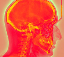
Computed Radiography (CR) uses very similar equipment to conventional radiography except that in place of a film to create the image, an imaging plate (IP) made of photostimulable phosphor is used. The imaging plate housed in a special cassette and placed under the body part or object to be examined and the x-ray exposure is made. Hence, instead of taking an exposed film into a darkroom for developing in chemical tanks or an automatic film processor, the imaging plate is run through a special laser scanner, or CR reader, that reads and digitizes the image. The digital image can then be viewed and enhanced using software that has functions very similar to other conventional digital image-processing software, such as contrast, brightness, filtration and zoom.
Differences from Direct Radiography
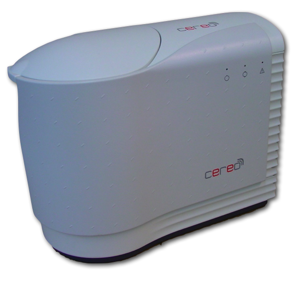
 CeReO - Computed Radiography Scanner
Computed radiography (CR) is often distinguished from Direct Radiography (DR). CR and DR have many similarities. Both CR and DR use a medium to capture x-ray energy and both produce a digital image that can be enhanced for soft copy diagnosis or further review. Both CR and DR can also present an image within seconds of exposure. CR generally involves the use of a cassette that houses the imaging plate, similar to traditional film-screen systems, to record the image whereas DR typically captures the image directly onto a flat panel detector without the use of a cassette. Image processing or enhancement can be applied on DR images as well as CR images due to the digital format of each. There are many different types of DR detectors in use in medicine and industry. Each type has its own merits and distinctions and may be applied to certain imaging requirements based on these attributes.
CR and DR should not be confused with fluoroscopy, where there is a continuous beam of radiation, and the images appear on the screen like on a TV. This is the system many people are familiar with, where the image of the article being x-rayed is viewed in real time on a monitor or display. Many people think airports use fluoroscopes for baggage inspection, when in fact LDAs (Linear Diode Arrays) are universally used to generate static images of luggage content. LDAs are also used in a wide variety of other screening and imaging applications, and are also presented in a digital format. Modern fluorosopes use a device called an image intensifier to enhance the analog output of the real time x-ray image, where it is picked up by either a video or CCD camera and digitally enhanced to reduce the noise inherent in the system.
Imaging plate
The CR imaging plate (IP) contains photostimulable storage phosphors, which store the radiation level received at each point in local electron energies. When the plate is put through the scanner, the scanning laser beam causes the electrons to relax to lower energy levels (photostimulated luminescence), emitting light that is detected by a photo-multiplier tube, which is then converted to an electronic signal. The electronic signal is then converted to discrete (digital) values and placed into the image processor pixel map.
Imaging plates can theoretically be re-used thousands of times if they are handled carefully. IP handling under industrial conditions, however, may result in damage after a few hundred uses. An image can be erased by simply exposing the plate to a room-level fluorescent light. Most laser scanners automatically erase the image plate after laser scanning is complete. The imaging plate can then be re-used. Reusable phosphor plates are environmentally safe but need to be disposed of according to local regulations.
Industrial applications
Common applications for computed radiography include:
- corrosion surveys on pipes, often through insulation;
- Examination of valves for erosion;
- Information shots on industrial components; e.g. checking to see if a valve is closed properly, or checking for obstructions in valves and pipes;
- Examination of boiler water walls;
- Weld examination for all AWS & ASME code applications including Sect. III & XI
- Automotive casting inspection
- High pressure braze joint inspection (aerospace)
- Wax pattern core integrity verification in investment casting foundries
- Best when used with Se75 for small core piping due to internal scatter created by Ir192's wavelength
- Code work for Nuclear Applications on all size piping
Medical applications
Computed Radiography systems are the most common in medical applications because they have proven reliability over more than two decades, flexibility to address a variety of clinical applications and lower costs to take multiple exam rooms digital. DR in the form of a portable detector starts at around $150,000.00, while a basic low volume CR can start as low as $30,000.00 (but higher volume, hospital-grade applications can be higher.) DR systems are generally sold as a full x-ray room replacements and tied to a single x-ray generator. CR IPs can be retrofitted to existing exam rooms and used in multiple x-ray sites since IPs are processed through a CR reader (scanner) that can be shared between multiple exam rooms.
Advantages
- No silver based film or chemicals are required to process film.
- Reduced film storage costs because images can be stored digitally.
- Computed radiography often requires fewer retakes due to under- or over-exposure which results in lower overall dose to the patient.
- Image acquisition is much faster - image previews can be available in less than 15 seconds.
- By adjusting image brightness and/or contrast, a wide range of thicknesses may be examined in one exposure, unlike conventional film based radiography, which may require a different exposure or multiple film speeds in one exposure to cover wide thickness range in a component.
- Images can be enhanced digitally to aid in interpretation.
- Images can be stored on disk or transmitted for off-site review.
- Ever growing technology makes the CR more affordable than ever today. With chemicals, dark room storage and staff to organize them, you could own a CR for the same monthly cost while being environmentally conscious, depending upon the size of the radiographic operation.
Disadvantages
- In medical applications, manual handling of the cassette housing the IP is considered a disadvantage versus DR but it also offers more flexibility for patient positioning.
- CR is still not an approved method for higher quality radiologic applications (aerospace), due to the possibility of digital manipulation to the captured image, the inherent geometric unsharpness and resultant lower spatial resolution as compared to film (radiographic) images, SNR (signal vs. noise) issues, sensitivity to scattered radiation, and the general lack of procedural consensus among primes and OEMs.
- There also are no quality (image resolution)standards for general radiography, only for mammography (21 CFR 900.12 (e)), however, competition among manufacturers has raised the bar and newer CR technologies with increased detective quantum efficiency (DQE) and higher spatial resolution have emerged.
- Imaging plates (IPs) are expensive and can be damaged if the system being used requires manual handling of the IPs. Theoretically, IPs may be reused thousands of times, but constant use will always result in damage to the IP and image artifacts, eventually to the point of necessary replacement.
Products
- Willick Engineering Company, Inc. - DÜRR NDT Products
- VMI Virtual Media Integradtion
- Oy. Ajat Ltd. CdTe CMOS imaging systems
- Viztek OPAL CR
- DÜRR DENTAL VistaScan Combi Plus
- DÜRR DENTAL VistaScan Perio Plus
- DÜRR DENTAL VistaScan Mini Plus
- DÜRR NDT HD-CR 35 NDT, HD-CR 43 NDT
- DÜRR MEDICAL CR 35 MED, CR 43 MED, CR 35 VET, CR 43 VET, CR 7 VET
- Hamamatsu Photonics (PMT)
- CONSORZIO REGIONALE MEDITEC
- Radlink CR PRO Series Systems
- Carestream Classic CR System (formerly Kodak)
- Carestream Elite CR System (formerly Kodak)
- Carestream Directview Max CR
- Carestream Vita CR
- Carestream Vita SE CR
- Carestream 975 / 950 / 900 / 850 / 825 / 800 CR System (formerly Kodak)
- Carestream Point of Care CR 360 / 140 / 120 (formerly Kodak)
- OREX ACL-4 / ACL-2
- CR-TECH CR 5020S
- FujiFilm FCR Capsula X
- FujiFilm FCR Capsula XL II
- FujiFilm FCR XG 5000
- FujiFilm FCR XG-1
- FujiFilm Profect CS (Mammography)
- FujiFilm Profect One (Mammography)
- iCRco 3600
- iCRco 3600sf
- Konica ImagePilot CR
- Konica Regius 190 / 170 / 150
- Konica Xpress CR
- Konica IQue CR
- Philips PCR Eleva S
- Agfa CR 30-X
- Agfa CR 35-X
- Agfa CR 75-X
- Agfa CR 85-X
- Agfa DX-S
- Agfa ADC Solo
See also
References
Categories:

Wikimedia Foundation. 2010.