- Sacroiliac joint
Infobox Anatomy
Name = Sacroiliac joint
Latin = articulatio sacroiliaca
GraySubject = 80
GrayPage = 306
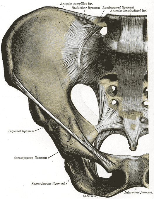
Caption = Articulations of pelvis. Anterior view.
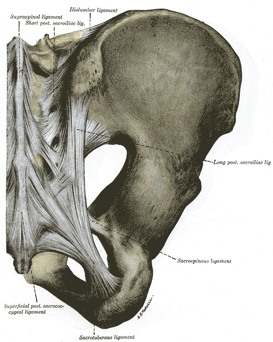
Caption2 = Articulations of pelvis. Posterior view.
System =
MeshName =
MeshNumber =
DorlandsPre = a_64
DorlandsSuf = 12161524
The sacroiliac joint is thejoint between thesacrum , at the base of the spine, and the ilium of thepelvis , which are joined byligaments . It is a strong, weightbearing synovial joint with irregular elevations and depressions that produce interlocking of the bones.Inflammation of this joint may be caused by "
sacroiliitis ", one cause of disablinglow back pain . With sacroiliitis, the individual may experience pain in the low back, buttocks and thighs, and may also have other symptoms of a rheumatic condition such as inflammation in the eyes orpsoriasis . Another condition of the sacroiliac joint is called "sacroiliac joint dysfunction" (also termed SI joint dysfunction). While SI joint dysfunction also causes low back and leg pain, and results from inflammation of the sacroiliac joint, it differs from sacroiliitis in that its origin is a disruption in the normal movement of the joint (too much or too little movement in the joint).Anatomy
The sacroiliac joints are 2 paired L-shaped synovial joints (joint permitting minimal motion) formed between the articular surfaces of the sacrum and ilium. The 2 sacroiliac joints move together as a unit and are considered bicondylar joints. The joints are covered by two different kinds cartilage; the sacral anterior edge has a hyaline cartilage and the ilium anterior edge a fibrocartilage. The stability of the SIJ's are maintained by various muscles and ligaments. As we age the characteristics of the sacroiliac joint change. The joint's surface remains flat until sometime after puberty. In our thirties and forties there is an increase in the size and number of elevations and depressions on the sacral and iliac surfaces.
Ligaments
*
Anterior sacroiliac ligament
*Interosseous sacroiliac ligament
*Posterior sacroiliac ligament Depending on the reference source cited, the anterior ligament may be described as just a thickening of the anterior joint capsule. The anterior ligament is certainly not as strong and well defined as are the posterior ligaments.The posterior sacroiliac (SI) ligaments can be further divided into short and long. There is a very strong structure which is called the dorsal interosseous ligament. This structure is stronger than bone; such that the pelvis will fracture before this structure tears. There is much we do not know regarding pelvic ligaments. For example it is known that ligaments become loose during pregnancy in response to hormones, especially relaxin; to allow widening of the joints during the birth process. We do not know if the interoseous membrane has the same type of receptors as the ligaments, and the specific ligament nerves (called mechanoreceptors/nociceptors) of the SI joint have not been studied in detail. The long and short SI ligaments can be palpated and the tone of the ligaments can be compared from one side of the body to the other.Physiology
The SI joints function include shock absorption for the spine through stretching in various directions and torque conversion, allowing the rotations that take place in the lower extremity to be transmitted up the spine. The SI joint may also provide a "self-locking" mechanism that helps you to walk. The joint locks on one side as weight is transferred from one leg to the other and through the pelvis the body weight is transmitted from the sacrum to the hip bone. These joints bear the weight of the twists and turns of the trunk of the body. It is common for the SI joint to become stiff and actually "lock" as we age.
The motions of the sacroiliac joint's are:
*Anterior innominate tilt
*Posterior innominate tilt
*Sacral flexion (or nutation)
*Sacral extension (or counter-nutation)ymptoms
*Mechanical SIJ dysfunction usually causes a dull ache.
*The pain may become worse and sharp whilst doing activities such as standing up from a seated position, or lifting the knee up to the chest during stair climbing.
*The pain in your buttocks and low back and will often radiate to the front into the groin.
*Typically the pain is felt in the low back and/or on the same side as the SIJ problem.
*Noticing frequent changes in body posture to avoid prolonged tension on the SIJ and ligaments.
*When SIJ dysfunction is severe, there can be referred ligament and joint pain into the hip, groin and leg.
*Pain can be referred from the SIJ down into the buttock or back of the thigh.
*Rib or mid back pain that increases with arm movement or prolonged sitting.
*Loss of bowel and/or bladder control.
*Pain during sexual intercourse.
*Occasionally there may be referred pain into the lower limb which can be mistaken for sciatica.
*Difficulty turning over in bed.Pregnancy
The hormonal changes of menstruation, pregnancy, and lactation can affect the integrity of the ligament support around the SIJ, which is why women often find the days leading up to their period are when the pain is at its worst. During pregnancy, female hormones are released that allow the connective tissues in the body to relax. The relaxation is necessary so that during delivery, the female pelvis can stretch enough to allow birth. This stretching results in changes to the SIJ's, making them hypermobile - extra or overly mobile. Over a period of years, these changes can eventually lead to wear-and-tear arthritis. As would be expected, the more pregnancies a woman has, the higher her chances of SI joint problems. During the pregnancy micro tears and small gas pocket can appear within the joint. Traumatic incidents, biomechanical mal-alignments and hormonal changes can all lead to SIJ dysfunction. The self-braced position of the SIJ can be altered by these factors and the joint can lose its stability. SIJ dysfunction puts abnormal pressures on the joint surfaces, ligaments and surrounding muscles. In some situations, pain can be felt at the front of the pelvis, down near the pubic bone. Also, when the front part of the pelvis moves down relative to the spine, it stretches the psoas muscle. The ligaments helping to stabilize the SIJ can become lax and this, together with increased load on the spine due to the pregnancy, can cause altered SI joint mechanics and pain. Any type of back or sacroiliac problem that causes excessive movement of the pelvis can result in excessive movement in the pubic symphysis and its ligaments. Sometimes an obvious limp is present due to one or both of the joints locking. There is a relation between asymmetric laxity of the sacroiliac joints and pregnancy related pelvic girdle pain. This condition can begin either pre or post partum. Women are eight to 10 times more likely to suffer from sacroiliac pain than men, mostly because of structural and hormonal differences between the sexes. Her anatomy allows one less sacral segment to lock with the pelvis and this influences instability.
Gillet's Test
The Sacroiliac joint dysfunction (SI Joint Dysfunction) can be tested with the Stork Test.
# With the patient standing and the examiner sitting behind, the examiner's left thumb is placed over the most posterior portion of the left posterior superior iliac spine (PSIS) and the right thumb overlying the midline of the sacrum at the same level.
# Examiner asks the patient to flex the left hip and knee to a minimum of 90 degrees of the hip flexion. Imagine making an "L" with the leg and thigh.
# A negative test finds the left thumb on the posterior superior iliac spine (PSIS) moving caudad (towards the tail) in relation to the right thumb on the sacrum.
# The thumb placements are reversed, and the patient is asked to raise the right leg in similar fashion.
# A positive finding occurs when the thumb on the PSIS moves cranially (towards the head) in relation to the thumb on the sacrum.
# The findings of this test are correlated with those of the standing flexion test. The Stork test is more specific for SI joint Restriction.
# If the patient has difficulty standing on one foot to perform the test, proprioceptive sensory motor balance deficit should be further evaluated.Other tests
Patrick's test ,Gaenslen's test andYeoman's test are widely-used tests forsacroiliitis .These tests have to be interpreted very cautiously. There are many more tests available and passive motion tests can be performed to evaluate motion going through the structure. These are called spring tests. The Hesch Method is an extensive evaluation and treatment system that utilizes many spring tests applied to various parts of the bony pelvis. These are performed with various positions such as having the person lie prone, supine, sitting, side-lying, prone extension and prone flexion (also called Muslim Prayer Position).
In popular culture
Because "sacroiliac" is a colloquially peculiar-sounding word, it often has been used for humorous or rhyming purposes in popular culture.
*In their
1975 old schoolFunk song Ride On, Parliament invites the listener to shake his sacroiliac.*In their
1981 New Wave song Rapture, the band Blondie mentions people dancing cheek to cheek, with their sacroiliacs touching.*In their
1982 old school hip-hop song The Message,Grandmaster Flash and The Furious Five , the singer mentions, "Can't stop to turn around, broke my sacroiliac"*In the 10th season episode of the animated television sitcom
King of the Hill titled "A Portrait of the Artist as a Young Clown," a comedy teacher instructs his class (including Bobby Hill) that "sacroiliac" is a "funny-sounding body part" because it has three "funny letters": two "k" sounds and an "oi" sound.*In the
1989 songLet Your Backbone Slide ,Maestro Fresh Wes claims "There's so many suckers on my sacroiliac/It's like a rap-sack, backpack."External links
*
*
Wikimedia Foundation. 2010.