- Crystallography
-
For the book of poetry, see Crystallography (book).
 A crystalline solid: atomic resolution image of strontium titanate. Brighter atoms are Sr and darker ones are Ti.
Crystallography is the experimental science of the arrangement of atoms in solids. The word "crystallography" derives from the Greek words crystallon = cold drop / frozen drop, with its meaning extending to all solids with some degree of transparency, and grapho = write.
Before the development of X-ray diffraction crystallography (see below), the study of crystals was based on their geometry. This involves measuring the angles of crystal faces relative to theoretical reference axes (crystallographic axes), and establishing the symmetry of the crystal in question. The former is carried out using a goniometer. The position in 3D space of each crystal face is plotted on a stereographic net, e.g. Wulff net or Lambert net. In fact, the pole to each face is plotted on the net. Each point is labelled with its Miller index. The final plot allows the symmetry of the crystal to be established.
Crystallographic methods now depend on the analysis of the diffraction patterns of a sample targeted by a beam of some type. Although X-rays are most commonly used, the beam is not always electromagnetic radiation. For some purposes electrons or neutrons are used. This is facilitated by the wave properties of the particles. Crystallographers often explicitly state the type of illumination used when referring to a method, as with the terms X-ray diffraction, neutron diffraction and electron diffraction.
These three types of radiation interact with the specimen in different ways. X-rays interact with the spatial distribution of the valence electrons, while electrons are charged particles and therefore feel the total charge distribution of both the atomic nuclei and the surrounding electrons. Neutrons are scattered by the atomic nuclei through the strong nuclear forces, but in addition, the magnetic moment of neutrons is non-zero. They are therefore also scattered by magnetic fields. When neutrons are scattered from hydrogen-containing materials, they produce diffraction patterns with high noise levels. However, the material can sometimes be treated to substitute hydrogen for deuterium. Because of these different forms of interaction, the three types of radiation are suitable for different crystallographic studies.
Contents
Theory
Condensed matter physics 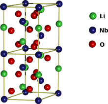
Phases · Phase transition Solid · Liquid · Gas · Bose-Einstein condensate · Fermionic condensate · Fermi gas · Fermi liquid · Supersolid · Superfluid · Luttinger liquidPhase phenomenaOrder parameter · Phase transitionElectronic phasesInsulator · Mott insulator · Semiconductor · Semimetal · Conductor · Superconductor · Thermoelectric · Piezoelectric · FerroelectricElectronic phenomenaMagnetic phasesGenerally, an image of a small object is made using a lens to focus the illuminating radiation, as is done with the rays of the visible spectrum in light microscopy. However, the wavelength of visible light (about 4000 to 7000 angstroms) is three orders of magnitude longer than the length of typical atomic bonds and atoms themselves (about 1 to 2 angstroms). Therefore, obtaining information about the spatial arrangement of atoms requires the use of radiation with shorter wavelengths, such as X-rays or neutrons beam. Employing shorter wavelengths implied abandoning microscopy and true imaging, however, because there exists no material from which a lens capable of focusing this type of radiation can be created. (That said, scientists have had some success focusing X-rays with microscopic Fresnel zone plates made from gold, and by critical-angle reflection inside long tapered capillaries.)[1] Diffracted x-ray or neutrons beams cannot be focused to produce images, so the sample structure must be reconstructed from the diffraction pattern. Sharp features in the diffraction pattern arise from periodic, repeating structure in the sample, which are often very strong due to coherent reflection of many photons from many regularly spaced instances of similar structure, while non-periodic components of the structure result in diffuse (and usually weak) diffraction features. Said more simply, areas with a higher density and repetition of atom order tend to reflect more light toward one point in space when compared to those areas with fewer atoms and less repetition.
Because of their highly ordered and repetitive structure, crystals give diffraction patterns of sharp Bragg reflection spots, and are ideal for analyzing the structure of solids.
Notation
Main article: Miller index- Coordinates in square brackets such as [100] denote a direction vector (in real space).
- Coordinates in angle brackets or chevrons such as <100> denote a family of directions which are related by symmetry operations. In the cubic crystal system for example, <100> would mean [100], [010], [001] or the negative of any of those directions.
- Miller indices in parentheses such as (100) denote a plane of the crystal structure, and regular repetitions of that plane with a particular spacing. In the cubic system, the normal to the (hkl) plane is the direction [hkl], but in lower-symmetry cases, the normal to (hkl) is not parallel to [hkl].
- Indices in curly brackets or braces such as {100} denote a family of planes and their normals which are equivalent in cubic materials due to symmetry operations, much the way angle brackets denote a family of directions. In non-cubic materials, <hkl> is not necessarily perpendicular to {hkl}.
Technique
Some materials studied using crystallography, proteins for example, do not occur naturally as crystals. Typically, such molecules are placed in solution and allowed to crystallize over days, weeks, or months through vapor diffusion. A drop of solution containing the molecule, buffer, and precipitants is sealed in a container with a reservoir containing a hygroscopic solution. Water in the drop diffuses to the reservoir, slowly increasing the concentration and allowing a crystal to form. If the concentration were to rise more quickly, the molecule would simply precipitate out of solution, resulting in disorderly granules rather than an orderly and hence usable crystal.
Once a crystal is obtained, data can be collected using a beam of radiation. Although many universities that engage in crystallographic research have their own X-ray producing equipment, synchrotrons are often used as X-ray sources, because of the purer and more complete patterns such sources can generate. Synchrotron sources also have a much higher intensity of X-ray beams, so data collection takes a fraction of the time normally necessary at weaker sources. Complementary neutron crystallography techniques are used to enhance hydrogen atoms positions. Such techniques are available in Neutron facilities.
Producing an image from a diffraction pattern requires sophisticated mathematics and often an iterative process of modelling and refinement. In this process, the mathematically predicted diffraction patterns of an hypothesized or "model" structure are compared to the actual pattern generated by the crystalline sample. Ideally, researchers make several initial guesses, which through refinement all converge on the same answer. Models are refined until their predicted patterns match to as great a degree as can be achieved without radical revision of the model. This is a painstaking process, made much easier today by computers.
The mathematical methods for the analysis of diffraction data only apply to patterns, which in turn result only when waves diffract from orderly arrays. Hence crystallography applies for the most part only to crystals, or to molecules which can be coaxed to crystallize for the sake of measurement. In spite of this, a certain amount of molecular information can be deduced from the patterns that are generated by fibers and powders, which while not as perfect as a solid crystal, may exhibit a degree of order. This level of order can be sufficient to deduce the structure of simple molecules, or to determine the coarse features of more complicated molecules. For example, the double-helical structure of DNA was deduced from an X-ray diffraction pattern that had been generated by a fibrous sample.
Crystallography in materials engineering
 An example of a cubic lattice
Crystallography is a tool that is often employed by materials scientists. In single crystals, the effects of the crystalline arrangement of atoms is often easy to see macroscopically, because the natural shapes of crystals reflect the atomic structure. In addition, physical properties are often controlled by crystalline defects. The understanding of crystal structures is an important prerequisite for understanding crystallographic defects. Mostly, materials do not occur in a single crystalline, but poly-crystalline form, such that the powder diffraction method plays a most important role in structural determination.
A number of other physical properties are linked to crystallography. For example, the minerals in clay form small, flat, platelike structures. Clay can be easily deformed because the platelike particles can slip along each other in the plane of the plates, yet remain strongly connected in the direction perpendicular to the plates. Such mechanisms can be studied by crystallographic texture measurements.
In another example, iron transforms from a body-centered cubic (bcc) structure to a face-centered cubic (fcc) structure called austenite when it is heated. The fcc structure is a close-packed structure, and the bcc structure is not, which explains why the volume of the iron decreases when this transformation occurs.
Crystallography is useful in phase identification. When performing any process on a material, it may be desired to find out what compounds and what phases are present in the material. Each phase has a characteristic arrangement of atoms. Techniques like X-ray or neutron diffraction can be used to identify which patterns are present in the material, and thus which compounds are present.
Crystallography covers the enumeration of the symmetry patterns which can be formed by atoms in a crystal and for this reason has a relation to group theory and geometry. See symmetry group.
Biology
X-ray crystallography is the primary method for determining the molecular conformations of biological macromolecules, particularly protein and nucleic acids such as DNA and RNA. In fact, the double-helical structure of DNA was deduced from crystallographic data. The first crystal structure of a macromolecule was solved in 1958 [2] A three-dimensional model of the myoglobin molecule obtained by X-ray analysis.[3] The Protein Data Bank (PDB) is a freely accessible repository for the structures of proteins and other biological macromolecules. Computer programs like RasMol or Pymol can be used to visualize biological molecular structures. Neutron crystallography is often used to help refine structures obtained by x-ray methods or to solve a specific bond; the methods are often viewed as complementary, as x-rays are sensitive to electron positions and scatter most strongly off heavy atoms, while neutrons are sensitive to nucleus positions and scatter strongly off many light isotopes, including hydrogen and deuterium. Electron crystallography has been used to determine some protein structures, most notably membrane proteins and viral capsids.
Scientists of note
- William Barlow
- John Desmond Bernal
- William Henry Bragg
- William Lawrence Bragg
- Auguste Bravais
- Martin Julian Buerger
- Francis Crick
- Pierre Curie
- Peter Debye
- Boris Delone
- Jack Dunitz
- Paul Peter Ewald
- Evgraf Stepanovich Fedorov
- Rosalind Franklin
- Georges Friedel
- Paul Heinrich von Groth
- René Just Haüy
- Carl Hermann
- Johann Friedrich Christian Hessel
- Dorothy Crowfoot Hodgkin
- Robert Huber
- Aaron Klug
- Max von Laue
- Kathleen Lonsdale
- Ernest-François Mallard
- Charles-Victor Mauguin
- William Hallowes Miller
- Friedrich Mohs
- Paul Niggli
- Arthur Lindo Patterson
- Max Perutz
- Hugo Rietveld
- Jean-Baptiste L. Romé de l'Isle
- Paul Scherrer
- Arthur Moritz Schönflies
- Dan Shechtman
- Tej P. Singh
- Constance Tipper
- Christian Samuel Weiss
- Don Craig Wiley
- Ralph Walter Graystone Wyckoff
- Ada Yonath
See also
- Atomic packing factor
- Condensed matter physics
- Crystal engineering
- Crystal growth
- Crystal optics
- Crystal system
- Crystal
- Crystallite
- Crystallization processes
- Crystallographic database
- Crystallographic group
- Dynamical theory of diffraction
- Electron crystallography
- Euclidean plane isometry
- Fixed points of isometry groups in Euclidean space
- Fractional coordinates
- Group action
- Laser-heated pedestal growth
- Materials science
- Metallurgy
- Mineralogy
- Neutron crystallography
- NMR crystallography
- Neutron diffraction at OPAL
- Neutron diffraction at the ILL
- Permutation group
- Point group
- Quasicrystal
- Solid state chemistry
- Space group
- Symmetric group
References
Further reading
- Burns, G.; Glazer, A.M. (1990). Space Groups for Scientists and Engineers (2nd ed.). Boston: Academic Press, Inc. ISBN 0-12-145761-3.
- Clegg, W (1998). Crystal Structure Determination (Oxford Chemistry Primer). Oxford: Oxford University Press. ISBN 0-19-855-901-1.
- Drenth, J (1999). Principles of Protein X-Ray Crystallography. New York: Springer-Verlag. ISBN 0-387-98587-5.
- Giacovazzo, C; Monaco HL, Viterbo D, Scordari F, Gilli G, Zanotti G, and Catti M (1992). Fundamentals of Crystallography. Oxford: Oxford University Press. ISBN 0-19-855578-4.
- Glusker, JP; Lewis M, Rossi M (1994). Crystal Structure Analysis for Chemists and Biologists. New York: VCH Publishers. ISBN 0-471-18543-4.
- O'Keeffe, M.; Hyde, B.G. (1996). Crystal Structures; I. Patterns and Symmetry. Washington, DC: Mineralogical Society of America, Monograph Series. ISBN 0-939950-40-5.
- Applied Computational Powder Diffraction Data Analysis
- Edited by R. A. Young (1993). Young, R.A.. ed. The Rietveld Method. Oxford: Oxford University Press & International Union of Crystallography. ISBN 0-19-855577-6.
External links
- American Crystallographic Association
- Learning Crystallography
- Crystal Lattice Structures
- Vega Science Trust Interviews on Crystallography Freeview video interviews with Max Pertuz, Rober Huber and Aaron Klug.
- Commission on Crystallographic Teaching, Pamphlets
- Ames Laboratory, US DOE Crystallography Research Resources
- International Union of Crystallography
Categories:- Chemistry
- Condensed matter physics
- Crystallography
- Instrumental analysis
- Materials science
- Neutron related techniques
- Synchrotron-related techniques


Wikimedia Foundation. 2010.