- Capsule of hip joint
Infobox Anatomy
Name = PAGENAME
Latin = capsula articularis coxae
GraySubject = 92
GrayPage = 334
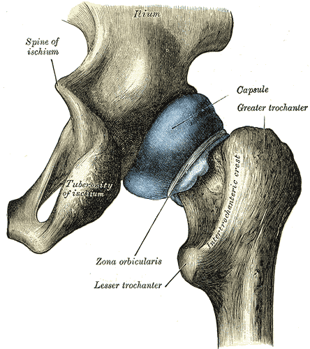
Caption = Capsule of hip-joint (distended). Posterior aspect.

Caption2 = Hip-joint, front view. The capsular ligament has been largely removed. (Capsular ligament visible at center.)
Precursor =
System =
Artery =
Vein =
Nerve =
Lymph =
MeshName =
MeshNumber =
DorlandsPre = c_07
DorlandsSuf = 12211231
The articular capsule (capsular ligament) is strong and dense.Above, it is attached to the margin of the
acetabulum 5 to 6 mm. beyond theglenoidal labrum behind; but in front, it is attached to the outer margin of the labrum, and, opposite to the notch where the margin of the cavity is deficient, it is connected to thetransverse ligament , and by a few fibers to the edge of theobturator foramen .It surrounds the
neck of the femur , and is attached, in front, to theintertrochanteric line ; above, to the base of the neck; behind, to the neck, about 1.25 cm. above theintertrochanteric crest ; below, to the lower part of the neck, close to the lesser trochanter.From its femoral attachment some of the fibers are reflected upward along the neck as longitudinal bands, termed "retinacula".
The capsule is much thicker at the upper and forepart of the joint, where the greatest amount of resistance is required; behind and below, it is thin and loose.
It consists of two sets of fibers, circular and longitudinal.
The circular fibers, "zona orbicularis", are most abundant at the lower and back part of the capsule, and form a sling or collar around the neck of the
femur .Anteriorly they blend with the deep surface of the
iliofemoral ligament , and gain an attachment to theanterior inferior iliac spine .The longitudinal fibers are greatest in amount at the upper and front part of the capsule, where they are reinforced by distinct bands, or accessory ligaments, of which the most important is the
iliofemoral ligament .The other accessory bands are known as the
pubocapsular and theischiocapsular ligament s.The external surface of the capsule is rough, covered by numerous muscles, and separated in front from the
Psoas major andIliacus by a bursa, which not infrequently communicates by a circular aperture with the cavity of the joint.
Wikimedia Foundation. 2010.