- Epidural space
Infobox Anatomy
Name = Epidural space
Latin = spatium extradurale
GraySubject = 193
GrayPage = 875
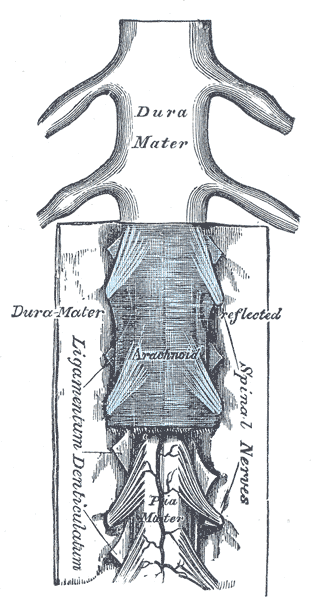
Caption = Themedulla spinalis and its membranes.
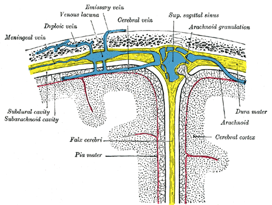
Caption2 = Diagrammatic representation of a section across the top of theskull , showing the membranes of the brain, etc.
System =
MeshName =
MeshNumber =
DorlandsPre = s_16
DorlandsSuf = 12746475
:"For a discussion about the anesthetic procedure, seeEpidural ."In the spine, the epidural space (also known as "extradural space" or "peridural space") is the outermost part of thespinal canal . It is the space within the canal (formed by the surrounding vertebrae) lying outside thedura mater (which encloses thearachnoid mater ,subarachnoid space , the cerebrospinal fluid, and thespinal cord ). In humans the epidural space containslymphatics , spinal nerve roots, loose fatty tissue, smallarteries , and a network of large, thin-walled blood vessels called theepidural venous plexus .In humans
The upper limit of the epidural space is the
foramen magnum , which is the point where the spine meets the base of theskull . The lower limit is at the tip of thesacrum , at thesacrococcygeal membrane .In the head, the dura is continuous with the
periosteum , the tough fibrous lining of the inside of theskull . This means that, in the head, the epidural space is known as a potential space, which means that normally it does not exist. In rare circumstances, a torn artery (e.g. themiddle meningeal artery ) may cause bleeding which is sufficient to separate both the dura andperiosteum from the bone; this is anepidural hematoma .The space between the dura and the arachnoid (in both head and spine), the
subdural space , is also a potential space. Bleeding may also occur here.In other mammals
In other mammals, the relationship between the spinal canal and its contents is similar to that in humans, although many species possess a tail into which the epidural space is prolonged.
A unique property of the epidural venous plexus is that the veins are prevented from collapsing due to external pressure because the bony spinal canal prevents that pressure being transmitted. This means that for many diving mammals, e.g.
whale s, when diving a large fraction of venous return to the heart takes place via the epidural space, as veins such as thevena cava may be substantially compressed by the pressure at depth.ee also
*
Subarachnoid space
*Subdural space
*Meninges External links
*
* [http://depts.washington.edu/anesth/regional/epiduralspaceframes.html "Anatomy of the Epidural Space" at washington.edu]
Wikimedia Foundation. 2010.