- Hydatidiform mole
-
Hydatidiform mole Classification and external resources 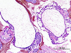
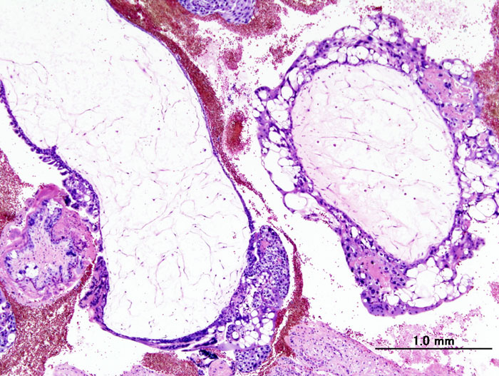
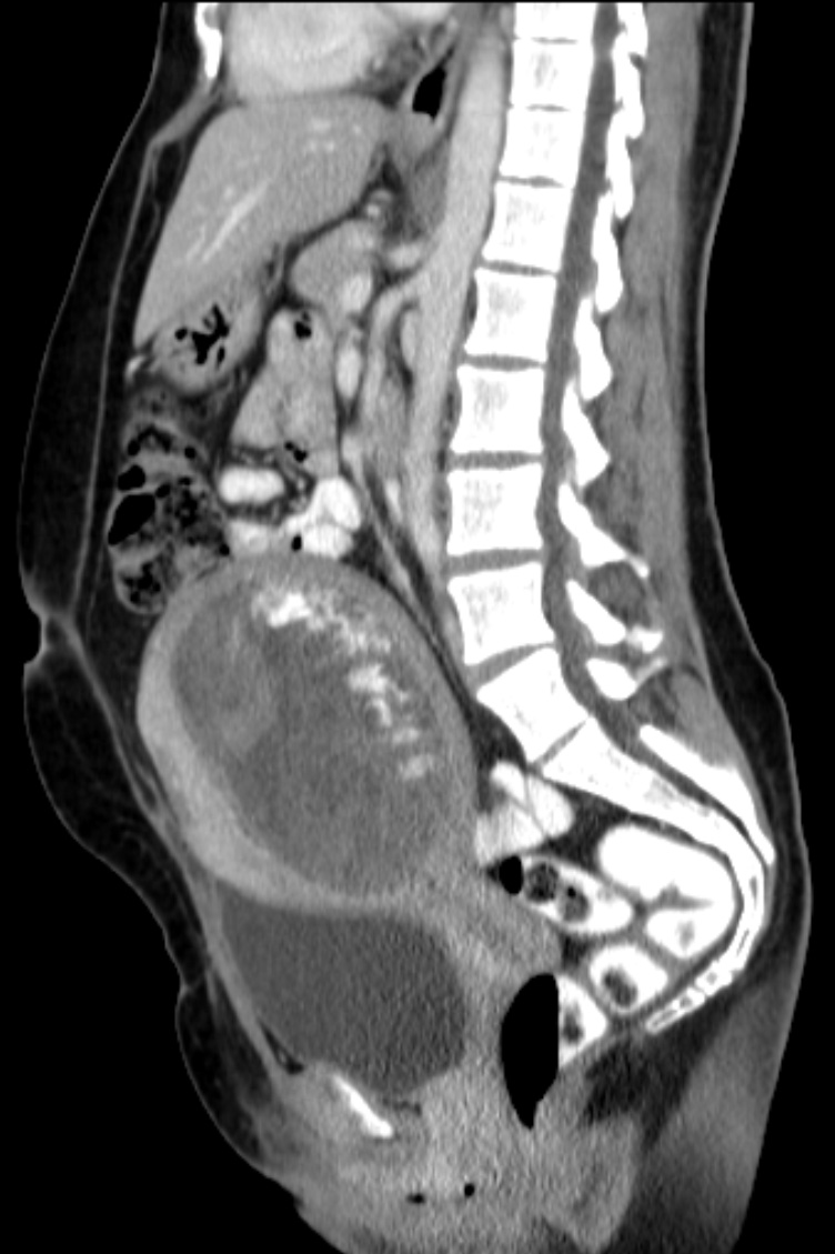
Histopathogic image of hydatidiform mole (complete type). H & E stain.ICD-10 O01, D39.2 ICD-9 630 ICD-O: M9100/0 OMIM 231090 DiseasesDB 6097 MedlinePlus 000909 eMedicine med/1047 med/866 MeSH D006828 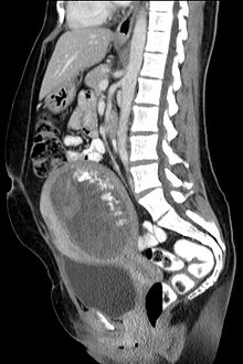 Hydatidiform mole on CT, sagittal view
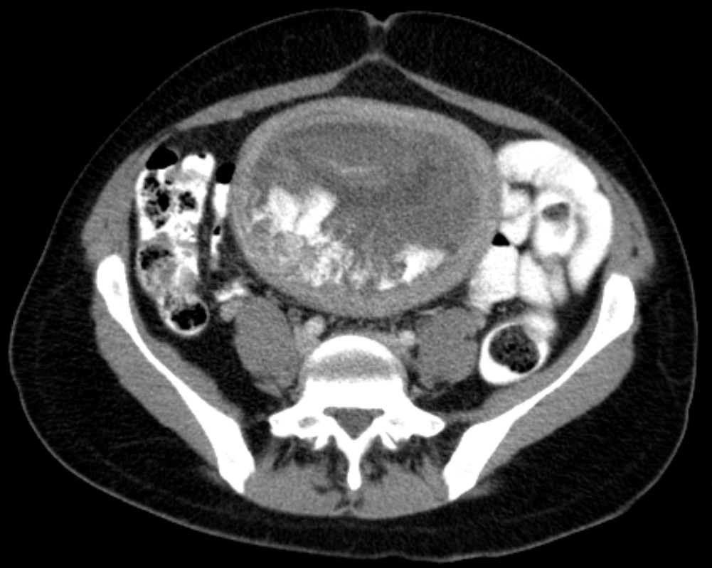
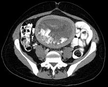 Hydatidiform mole on CT, axial view
Molar pregnancy is an abnormal form of pregnancy, wherein a non-viable, fertilized egg implants in the uterus, and thereby converts normal pregnancy processes into pathological ones. It is characterized by the presence of a hydatidiform mole (or hydatid mole, mola hydatidosa).[1] Molar pregnancies are categorized into partial and complete moles.
A complete mole is caused by a single sperm combining with an egg which has lost its DNA (the sperm then reduplicates forming a "complete" 46 chromosome set) [2] The genotype is typically 46,XX (diploid) due to subsequent mitosis of the fertilizing sperm, but can also be 46,XY (diploid).[2] In contrast, a partial mole occurs when an egg is fertilized by two sperm or by one sperm which reduplicates itself yielding the genotypes of 69,XXY (triploid) or 92,XXXY (quadraploid).[2] Complete hydatidiform moles have a higher risk of developing into choriocarcinoma — a malignant tumor of trophoblast cells — than do partial moles.
The etymology is derived from hydatisia (Greek "a drop of water"), referring to the watery contents of the cysts, and mole (from Latin mola = millstone/false conception).[3] The term, however, comes from the similar appearance of the cyst to a hydatid cyst in an Echinococcosis.[4]
A hydatidiform mole conception may be categorized in medical terms as one type of non-induced (natural) "missed abortion".[5] - referred to colloquially as a "missed miscarriage", because the pregnancy has become non-viable (miscarried) but was not immediately expelled (therefore was "missed").
Contents
Natural history
A hydatidiform mole is a pregnancy/conceptus in which the placenta contains grapelike vesicles (small sacs) that are usually visible with the naked eye. The vesicles arise by distention of the chorionic villi by fluid. When inspected in the microscope, hyperplasia of the trophoblastic tissue is noted. If left untreated, a hydatidiform mole will almost always end as a spontaneous abortion (miscarriage).
Based on morphology, hydatidiform moles can be divided into two types: In complete moles, all the chorionic villi are vesicular, and no sign of embryonic or fetal development is present. In partial moles some villi are vesicular, whereas others appear more normal, and embryonic/fetal development may be seen but the fetus is always malformed and is never viable.
Hydatidiform moles are a common complication of pregnancy, occurring once in every 1000 pregnancies in the US, with much higher rates in Asia (e.g. up to one in 100 pregnancies in Indonesia).[6]
In rare cases a hydatidiform mole co-exists in the uterus with a normal, viable fetus. These cases are due to twinning. The uterus contains two conceptuses: one with an abnormal placenta and no viable fetus (the mole), and one with a normal placenta and a viable fetus. Under careful surveillance it is often possible for the woman to give birth to the normal child and to be cured of the mole.[7]
The etiology of this condition is not completely understood. Potential risk factors may include defects in the egg, abnormalities within the uterus, or nutritional deficiencies. Women under 20 or over 40 years of age have a higher risk. Other risk factors include diets low in protein, folic acid, and carotene.[8] The diploid set of sperm-only DNA means that all chromosomes have sperm-patterned methylation suppression of genes. This leads to overgrowth of the syncytiotrophoblast whereas dual egg-patterned methylation leads to a devotion of resources to the embryo, with an underdeveloped syncytiotrophoblast. This is considered to be the result of evolutionary competition with male genes driving for high investment into the fetus versus female genes driving for resource restriction to maximise the number of children.[9]
Parental origin
In most complete moles, all nuclear genes are inherited from the father only (androgenesis). In approximately 80% of these androgenetic moles, the most probable mechanism is that an empty egg is fertilized by a single sperm, followed by a duplication of all chromosomes/genes (a process called "endoreduplication"). In approximately 20% of complete moles the most probable mechanism is that an empty egg is fertilised by two sperm. In both cases, the moles are diploid (i.e. there are two copies of every chromosome). In all these cases, the mitochondrial genes are inherited from the mother, as usual.
Most partial moles are triploid (three chromosome sets). The nucleus contains one maternal set of genes and two paternal sets. The mechanism is usually the reduplication of the paternal haploid set from a single sperm, but may also be the consequence of dispermic (two sperm) fertilization of the egg.[10]
In rare cases, hydatidiform moles are tetraploid (four chromosome sets) or have other chromosome abnormalities.
A small percentage of hydatidiform moles have biparental diploid genomes, as in normal living persons; they have two sets of chromosomes, one inherited from each biological parent. Some of these moles occur in women who carry mutations in the gene NLRP7, predisposing them towards molar pregnancy. These rare variants of hydatidiform mole may be complete or partial.[11][12][13]
Clinical presentation and diagnosis
Molar pregnancies usually present with painless vaginal bleeding in the fourth to fifth month of pregnancy.[1] The uterus may be larger than expected, or the ovaries may be enlarged. There may also be more vomiting than would be expected (hyperemesis). Sometimes there is an increase in blood pressure along with protein in the urine. Blood tests will show very high levels of human chorionic gonadotropin (hCG).[14]
The diagnosis is strongly suggested by ultrasound (sonogram), but definitive diagnosis requires histopathological examination. On ultrasound, the mole resembles a bunch of grapes ("cluster of grapes" or "honeycombed uterus" or "snow-storm"[15]). There is increased trophoblast proliferation and enlarging of the chorionic villi.[16] Angiogenesis in the trophoblasts is impaired as well.[16]
Sometimes symptoms of hyperthyroidism are seen, due to the extremely high levels of hCG, which can mimic the normal Thyroid-stimulating hormone (TSH).[14]
Treatment
Hydatidiform moles should be treated by evacuating the uterus by uterine suction or by surgical curettage as soon as possible after diagnosis, in order to avoid the risks of choriocarcinoma.[17] Patients are followed up until their serum human chorionic gonadotrophin (hCG) level has fallen to an undetectable level. Invasive or metastatic moles (cancer) may require chemotherapy and often respond well to methotrexate. The response to treatment is nearly 100%. Patients are advised not to conceive for one year after a molar pregnancy. The chances of having another molar pregnancy are approximately 1%.
Management is more complicated when the mole occurs together with one or more normal fetuses.
Carboprost medication may be used to contract the uterus.
Prognosis
More than 80% of hydatidiform moles are benign. The outcome after treatment is usually excellent. Close follow-up is essential. Highly effective means of contraception are recommended to avoid pregnancy for at least 6 to 12 months.
In 10 to 15% of cases, hydatidiform moles may develop into invasive moles. This condition is named persistent trophoblastic disease (PTD). The moles may intrude so far into the uterine wall that hemorrhage or other complications develop. It is for this reason that a post-operative full abdominal and chest x-ray will often be requested.
In 2 to 3% of cases, hydatidiform moles may develop into choriocarcinoma, which is a malignant, rapidly-growing, and metastatic (spreading) form of cancer. Despite these factors which normally indicate a poor prognosis, the rate of cure after treatment with chemotherapy is high.
Over 90% of women with malignant, non-spreading cancer are able to survive and retain their ability to conceive and bear children. In those with metastatic (spreading) cancer, remission remains at 75 to 85%, although their childbearing ability is usually lost.
See also
References
- ^ a b Cotran RS, Kumar V, Fausto N, Nelso F, Robbins SL, Abbas AK (2005). Robbins and Cotran pathologic basis of disease (7th ed.). St. Louis, Mo: Elsevier Saunders. p. 1110. ISBN 0-7216-0187-1.
- ^ a b c Kumar, Vinay, ed (2010). Pathologic Basis of Disease (8th ed.). Saunders Elsevier. pp. 1057–1058. ISBN 1437707920.
- ^ Entries HYDATID n. (a.) and MOLE, n.6 in the Oxford English Dictionary online. (http://dictionary.oed.com/ — subscription required.)
- ^ http://medical-dictionary.thefreedictionary.com/hydatidiform
- ^ . PMID 2644599.
- ^ Di Cintio E, Parazzini F, Rosa C, Chatenoud L, Benzi G (1997). "The epidemiology of gestational trophoblastic disease". Gen Diagn Pathol 143 (2-3): 103–8. PMID 9443567.
- ^ Sebire NJ, Foskett M, Paradinas FJ, et al. (June 2002). "Outcome of twin pregnancies with complete hydatidiform mole and healthy co-twin". Lancet 359 (9324): 2165–6. doi:10.1016/S0140-6736(02)09085-2. PMID 12090984.
- ^ MedlinePlus Encyclopedia Hydatidiform mole
- ^ Paoloni-Giacobino A. (2007). "Epigenetics in reproductive medicine". Paediatr Res. May 61 (5 Pt 2): 51R–57R. doi:10.1203/pdr.0b013e318039d978. PMID 17413849.
- ^ Monga, Ash, ed (2006). Gynaecology by Ten Teachers (18th ed.). Hodder Arnold. pp. 99–101. ISBN 0340816627.
- ^ Lawler SD, Fisher RA, Dent J (May 1991). "A prospective genetic study of complete and partial hydatidiform moles". Am J Obstet Gynecol. 164 (5 Pt 1): 1270–7. PMID 1674641.
- ^ Wallace DC, Surti U, Adams CW, Szulman AE (1982). "Complete moles have paternal chromosomes but maternal mitochondrial DNA". Human genetics 61 (2): 145–7. doi:10.1007/BF00274205. PMID 6290372. http://www.springerlink.com/link.asp?id=w2555w4k4805h4h9.
- ^ Slim R, Mehio A (January 2007). "The genetics of hydatidiform moles: new lights on an ancient disease". Clin Genet. 71 (1): 25–34. doi:10.1111/j.1399-0004.2006.00697.x. PMID 17204043. Review.
- ^ a b Ganong WF, McPhee SJ, Lingappa VR (2005). Pathophysiology of Disease: An Introduction to Clinical Medicine (Lange). McGraw-Hill Medical. p. 639. ISBN 0-07-144159-X.
- ^ Woo J, Hsu C, Fung L, Ma H (1983). "Partial hydatidiform mole: ultrasonographic features". Aust N Z J Obstet Gynaecol 23 (2): 103–7. doi:10.1111/j.1479-828X.1983.tb00174.x. PMID 6578773.
- ^ a b Oslo University - Eksamensoppgaver i barnesykdommer; 9
- ^ Cotran RS, Kumar V, Fausto N, Nelso F, Robbins SL, Abbas AK (2005). Robbins and Cotran pathologic basis of disease (7th ed.). St. Louis, Mo: Elsevier Saunders. p. 1112. ISBN 0-7216-0187-1.
External links
- Humpath #3186 (Pathology images)
- Original source: MedlinePlus Encyclopedia Hydatidiform mole
- Clinically reviewed molar pregnancy and choriocarcinoma information for patients from Cancer Research UK
Germ cell tumors (ICD-O 9060–9119) (C45–C49/D17–D21, 171/214–215) Germinomatous Nongerminomatous Trophoblastic neoplasm: Gestational trophoblastic disease (Hydatidiform mole) · Choriocarcinoma · Placental site trophoblastic tumorCategories:- Germ cell neoplasia
- Pathology of pregnancy, childbirth and the puerperium
- Pregnancy with abortive outcome
Wikimedia Foundation. 2010.