- Somite
-
Somite 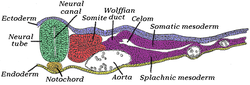
Transverse section of half of a chick embryo of forty-five hours' incubation. The dorsal (back) surface of the embryo is towards the top of this page, while the ventral (front) surface is towards the bottom. 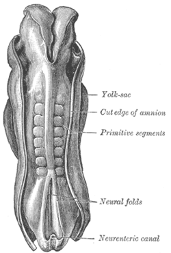
Dorsum of human embryo, 2.11 mm. in length. (The older term 'primitive segments' is used to identify the somites.) Latin somitus Gray's subject #9 52 Carnegie stage 9 Days 20[1] Precursor paraxial mesoderm Gives rise to dermatome, myotome, sclerotome Code TE E5.0.2.2.2.0.3 MeSH Somites A somite (or primitive segment in older texts) is a division of the body of an animal. In vertebrates this is mainly discernible in the embryo stage; in arthropods it is a characteristic of a hypothetical ancestor.
Contents
In vertebrates
In the developing vertebrate embryo, somites are masses of mesoderm distributed along the two sides of the neural tube and that will eventually become dermis (dermatome), skeletal muscle (myotome), and vertebrae (sclerotome).
Because the sclerotome differentiates before the other two structures, the term "dermomyotome" is sometimes used to describe the combined dermatome and myotome.[2]
Somites and their derivatives
Overview
The mesoderm forms at the same time as ectoderm and endoderm. The mesoderm that is lateral to (at the side of) the neural tube is called paraxial mesoderm. It is distinct from the mesoderm underneath the neural tube which is called the chordamesoderm and becomes the notochord. The paraxial mesoderm is initially called the “segmental plate” in the chick embryo or the “unsegmented mesoderm” in other vertebrates. As the primitive streak regresses and neural folds gather (to eventually become the neural tube), the paraxial mesoderm separates into blocks called somites. These somites have four compartments: the sclerotome forms the vertebrae and the rib cartilage; the myotome forms the musculature of the back, the ribs and the limbs; the dermatome forms the skin on the back; and the syndetome forms the tendons and some blood vessels. In addition the somites specify the migration paths of neural crest cells and spinal nerve axons.
Formation of somites
The cells which will become somites are committed before mesoderm becomes capable of forming somites. The formation of somites is called "somitogenesis". Somitogenesis depends on a clock and wave mechanism. Ocsilating Notch and Wnt signaling pathways provide the clock. The wave is a gradient of the FGF protein that is rostral to caudal (nose to tail gradient). Somites form one after the other down the length of the embryo from the head to the tail, with each new somite forming on the caudal (tail) side of those already in existing somites.
The timing of the interval is not universal. Different species have different interval timing. In the chick embryo somites are formed every 90 minutes. In the mouse the interval is variable.
-
- Sometimes called somitomeres, indicating a lack of complete separation between segments
- The outer cells become epithelium
- Inner cells remain as mesenchyme
- Number of somites can be used to determine what stage of development the embryo is at (because rates of development can be affected by temperature or other factors, absolute age is not a good indicator of development)
- Somites appear on both sides of the neural tube simultaneously
- Flipping stuff around has no effect on which ends develop as rostral or caudal, even fully excising the tissue has no effect, it will still order itself properly and at the right times
- Somite formation can be induced by Noggin-secreting cells
- Number of somites is species dependent and independent of embryo size (changed via surgery or genetic engineering)
- Chick: 50
- Mice: 65
- Snake: 500
Notch signalling
- Notch forms the boundaries of the somites
- DLL1 and DLL3 are Notch ligands, mutations of which cause various defects
- Notch regulates Hairy1, which sets up the caudal half of the somite
- Notch activation turns on Lunatic Fringe which inhibits the Notch receptor
- Notch activation also turns on HES which inactivates Lunatic Fringe, re-enabling the notch receptor, accounting for the oscillating clock model
- MESP2 induces EPHA4 which causes repulsive interaction that separates somites (causes segmentation)
- EPHA4 is restricted to the boundaries of somites
- EPHB2 also important for boundaries
Epithelialization of somites
- Fibronectin and N-cadherin are key to epithelialization
- Probably regulated by paraxis and MESP2
- MESP2 is regulated by Notch signaling
- Paraxis is regulated by processes involving the cytoskeleton
Anterior-posterior axis specification
- Hox genes specify which somite becomes which feature
- Specification occurs very early
- After somites are made, they are set for what they will become, transplantation of somites results in the wrong type of vertebrae forming in the wrong place
- The internal cells of the somite are not predestined
Somites produce
- Rib and vertebrae cartilage
- Muscles (through MyoD protein formation):
- Rib cage
- Limbs
- Abdominal wall
- Back and tongue
- Dorsal skin dermis (back skin)
In crustaceans
In crustacean biology, a somite is a segment of the hypothetical primitive crustacean body plan. In current crustaceans, several of those somites may be fused.
References
- ^ "The Third Week Of Life:". http://staff.um.edu.mt/acus1/Gasrtrulation.htm. Retrieved 2007-10-13.
- ^ "unc". http://www.med.unc.edu/embryo_images/unit-mslimb/mslimb_htms/mslimb008.htm. Retrieved 2007-10-19.
Additional images
-

Chick embryo of thirty-three hours’ incubation, viewed from the dorsal aspect. X 30.
External links
Developmental biology > Human embryogenesis (development of embryo) and development of fetus (TE E2.0) First three
weeksWeek 1Fertilization · Oocyte activation · Zygote · Cleavage · Morula · Blastula (Blastomere) · Blastocyst · Inner cell massWeek 2
(Bilaminar)Week 3
(Trilaminar)Archenteron/Primitive streak (Primitive pit, Primitive knot/Blastopore, Primitive groove) · Gastrula/Gastrulation · Regional specification · Embryonic discSplanchnopleuric mesenchymeChorda- · Paraxial (Somite/Somitomere) · Intermediate · Lateral plate (Intraembryonic coelom, Splanchnopleuric mesenchyme/Somatopleuric mesenchyme)Categories: -


Wikimedia Foundation. 2010.