- Rectus abdominis muscle
-
Rectus abdominis 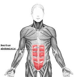
The human rectus abdominis muscle. Latin musculus rectus abdominis Gray's subject #118 415 Origin pubis Insertion Costal cartilage of ribs 5-7, xiphoid process of sternum Artery inferior epigastric artery Nerve segmentally by thoraco-abdominal nerves (T7 to T12) Actions Flexion of the lumbar spine Antagonist Erector spinae The rectus abdominis muscle, also known as the "6-pack," is a paired muscle running vertically on each side of the anterior wall of the human abdomen (and in some other animals). There are two parallel muscles, separated by a midline band of connective tissue called the linea alba (white line). It extends from the pubic symphysis/pubic crest inferiorly to the xiphisternum/xiphoid process and lower costal cartilages (5–7) superiorly.
It is contained in the rectus sheath.
The rectus is usually crossed by three fibrous bands linked by the tendinous intersections. While the "sixpack" is by far the most common configuration of the muscle bellies of the rectus, there exist rare anatomic variations which result in the appearance of eight ("eightpack"), ten, or asymmetrically arranged segments[citation needed]. All these variations are functionally equivalent.
 Rectus abdominis sheath
Contents
Function
The rectus abdominis is an important postural muscle. It is responsible for flexing the lumbar spine, as when doing a "crunch". The rib cage is brought up to where the pelvis is when the pelvis is fixed, or the pelvis can be brought towards the rib cage (posterior pelvic tilt) when the rib cage is fixed, such as in a leg-hip raise. The two can also be brought together simultaneously when neither is fixed in space.
The rectus abdominis assists with breathing and plays an important role in respiration in the event the patient is short of breath.[clarification needed] It also helps in keeping the internal organs intact and in creating intra-abdominal pressure, such as when exercising or lifting heavy weights, during forceful defecation or parturition (childbirth).
Blood supply
The rectus abdominis has several sources of arterial blood supply. In reconstructive surgery terms, it is a Mathes and Nahai[1] Type III muscle with 2 dominant pedicles. First, the inferior epigastric artery and vein (or veins) run superiorly on the posterior surface of the rectus abdominis, enter the rectus fascia at the arcuate line, and serve the lower part of the muscle. Second, the superior epigastric artery, a terminal branch of the internal thoracic artery, supplies blood to the upper portion. Finally, numerous small segmental contributions come from the lower six intercostal arteries as well.
Innervation
The muscles are innervated by thoracoabdominal nerves, which pierce the anterior layer of the rectus sheath.
Location
The rectus abdominis is a long flat muscle, which extends along the whole length of the front of the abdomen, and is separated from its fellow of the opposite side by the linea alba. The muscle is inserted by three portions of unequal size into the cartilages of the fifth, sixth, and seventh ribs. The upper portion, attached principally to the cartilage of the fifth rib, usually has some fibers of insertion into the anterior extremity of the rib itself.
Some fibers are occasionally connected with the costoxiphoid ligaments, and the side of the xiphoid process.
Damage
An abdominal muscle strain, also called a pulled abdominal muscle, is an injury to one of the muscles of the abdominal wall. A muscle strain occurs when the muscle is stretched too far. When this occurs the muscle fibers are torn. Most commonly, a strain causes microscopic tears within the muscle, but occasionally, in severe injuries, the muscle can rupture from its attachment.
Animals
 A feline rectus abdominis muscle, from a common housecat. This specimen has some remaining fascia and also shows the external obliques.
The rectus abdominis is similar in most vertebrates. The most obvious difference between animal and human abdominal musculature is that in animals, there are a different number of tendinous intersections.
Additional images
-

Anterior surface of sternum and costal cartilages
-

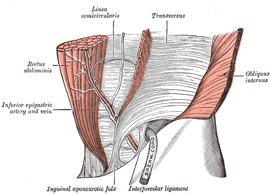
The interfoveolar ligament, seen from in front
-
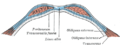
Diagram of sheath of rectus
-
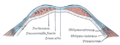
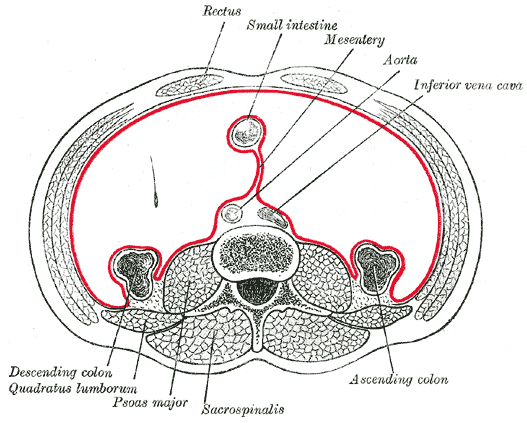
Diagram of a transverse section through the anterior abdomina wall, below the linea semicircularis
-
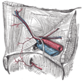
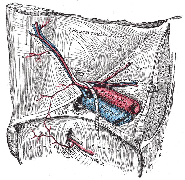
The relations of the femoral and abdominal inguinal rings, seen from within the abdomen. Right side
-

Horizontal disposition of the peritoneum in the lower part of the abdomen
-
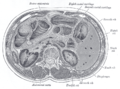
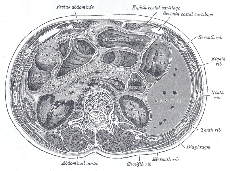
Transverse section through the middle of the first lumbar vertebra, showing the relations of the pancreas
-

The left side of the thorax
-

Surface anatomy of the front of the thorax and abdomen
-

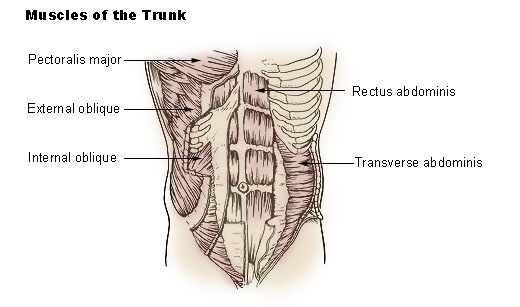
Muscles of the trunk
See also
References
- ^ Mathes SJ, Nahai F. Classification of the vascular anatomy of muscles: experimental and clinical correlation. Plast Reconstr Surg. Feb 1981;67(2):177-87.
External links
- LUC reca
- 168165454 at GPnotebook
- SUNY Figs 04:04-07 – "Muscles of the anterior chest wall with the pectoralis major muscles removed."
- SUNY Labs 18:01-0115 – "Thoracic Wall: The Anterior Thoracic Wall"
- SUNY Figs 35:06-07 – "Incision and reflection of the external abdominal oblique muscle."
- SUNY Figs 35:07-01 – "Incision and reflection of the internal abdominal oblique muscle."
- SUNY Labs 35:10-0100 – "Anterior Abdominal Wall: The Rectus Abdominis Muscle"
- Cross section at UV pembody/body12a
- Rectus+abdominis+muscle at eMedicine Dictionary
- Roche Lexicon - illustrated navigator, at Elsevier 25466.180-1
List of muscles of abdominopelvic cavity (TA A04.5, GA 4.408) Abdomen/
wallAnterior/
lateralMuscleFasciaFascia/abdominal fascia: panniculus adiposus (Fascia of Camper) · stratum membranosum (Fascia of Scarpa) · Transversalis fascia (Interfoveolar ligament)
Linea alba · Linea semilunaris · Inguinal triangle
Inguinal canal (Deep inguinal ring, Superficial inguinal ring, Intercrural fibers, Crura of superficial inguinal ring)
Inguinal ligament (Pectineal ligament, Lacunar ligament, Reflected ligament)PosteriorMuscleFasciaPelvis MuscleFasciafascia/pelvic fascia visceral (Rectovaginal fascia, Rectoprostatic fascia) · parietal (Obturator fascia/Tendinous arch, Piriformis fascia)
floor/diaphragm: Superior fascia of pelvic diaphragm (Pubovesical ligament, Puboprostatic ligament) · Inferior fascia of pelvic diaphragm
Anococcygeal bodyCategories:- Muscles of the torso
- Spine flexors
-


Wikimedia Foundation. 2010.