- Heart chamber
-
Heart chamber is a general term used to refer to any chambers of the mammalian heart. The heart consists of four chambers: the right and left atrium and the right and left ventricle. The top chambers are connected to the bottom chambers by valves and are separated by the coronary sulcus. The left and right side of the heart are separated by the posterior interventricular sulcus.
Contents
Function and Location
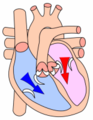
 This diagram shows blood flow through the different chambers of the heart. Blue arrows represent de-oxygenated blood and red arrows represent oxygenated blood.
- Right atrium: Situated in the upper right section of the heart, this chamber receives oxygen-depleted blood from the body via two major veins, the superior vena cava and the inferior vena cava. This chamber then pumps blood through the tricuspid valve into the right ventricle situated below.
- Right ventricle: Located below the right atrium, this chamber receives oxygen-depleted blood from the right atrium and pumps it through the pulmonary valve and into the lungs via the right and left pulmonary artery.
- Left atrium: This chamber sits opposite the right atrium and is the upper part of the heart that receives oxygen-rich blood from the lungs via the right and left pulmonary veins and pumps it through the bicuspid valve or mitral valve into the left ventricle.
- Left ventricle: This chamber is the lower part of the heart that receives oxygen-rich blood from the left atrium above it, and pumps it through the aortic valve to be distributed throughout the entire body via the aorta, including to the heart muscle itself through the coronary arteries.
The right atrium makes up the majority of the right boarder of the heart and the right ventricle makes up the majority of the inferior boarder. The left boarder of the heart consists of the left ventricle. In addition, the heart chamber located in the most superior position is the left atrium. The bottom left region, which includes the apex of the heart, sticks out more anteriorly compared to the rest of the heart.
Valves
Each heart chamber includes valves vital to the efficiency of the circulatory system. These valves open to allow the blood to flow in the right direction into, within, or out of the heart, and close to prevent the backflow of blood. There are four main valves that allow the chambers of the heart to carry out their function efficiently and they relax and contract during a heartbeat.
The Tricuspid Valve
- The tricuspid valve is located between the right atrium and the right ventricle. Blood passively fills the right atrium during diastole phase of the right atrium. When the right atrium is in systole phase, the contraction forces this valve open, thereby forcing any additional de-oxygenated blood into the ventricle.
The Semi-Lunar Pulmonary Valve
- The semi-lunar pulmonary valve is located between the right ventricle and the pulmonary trunk. During ventricle systole, the pressure within the chamber will increase until the valve is forced open. De-oxygenated blood can thereby be carried through the pulmonary arteries to each lung. This valve is named because the shape resembles half-moon pockets. When the ventricle relaxes, the pressure within the chamber decreases and blood fills these pockets causing them to close. [1]
The Mitral Valve
- The mitral valve is located between the left atrium and the left ventricle. Similar to the tricuspid valve, during the atrium's systole phase, the valve is forced open to allow the oxygenated blood from the lungs to enter into the left ventricle.
The Semi-Lunar Aortic Valve
- The semi-lunar aortic valve is located at the exit of the aorta and the left ventricle. The function of this valve mirrors that of the semi-lunar pulmonary valve, with one exception. Once the pressure in the left ventricle falls back during relaxation phase, oxygenated blood will fill up in the cusps of the semi-lunar aortic valve eventually causing them to close. This blood is what supplies the right and left coronary arteries which distribute blood to the muscle tissues of the heart. [2]
Ventricle Structure
The two ventricles are separated by a septum into the left ventricle and the right ventricle. [3] The ventricles of the heart are muscular chambers because they are responsible for propelling blood out of the heart.[4] The left ventricle is the thickest of the four chambers because it is the final chamber that oxygenated blood must be pumped through before it is distributed throughout the body via the circulatory system. The ventricles are also larger in size compared to the atria.
Atria Structure
The right atrium contains the sinoatrial node which sends an impulse throughout the heart causing the cardiac muscle of the atrium to contract in a coordinated, wave-like manner. [5] Since the atria are thin-walled and much less muscular than the ventricles, they are able to easily expand with the addition of blood. The forces of contraction are much weaker within the atria as a result. The contraction is strong enough, however to force 1/4 of the total blood volume entering into the ventricles.[6] The rest of the blood enters the atria passively.
Image Gallery
-

Another illustration of the human heart.
-
The heart ventricles in "diastole" or relaxtion phase.
-

The heart ventricles in "systole" or contraction phase.
-

An anterior view of the heart chambers.
-

Another illustration to show the direction of blood flow and the related valves.
See also
References
- ^ Martini, F.H., Timmons, M.J., & Tallitsch, R. B. (2012). Human Anatomy (7,th ed.). United States of America: Pearson Education Inc.
- ^ Martini, F.H., Timmons, M.J., & Tallitsch, R. B. (2012). Human Anatomy (7,th ed.). United States of America: Pearson Education Inc.
- ^ Baily, R. Ventricles of the Heart. retrieved from http://biology.about.com/od/anatomy/ss/ventricles.htm
- ^ Fogoros, R.N., (2003). The Hearts Chambers and Valves. retrieved from http://heartdisease.about.com/cs/starthere/a/chambersvalves.htm
- ^ Heart Anatomy. Retrieved from http://www.cardioconsult.com/Anatomy/
- ^ Sivasubramanian, M., (1997). The Atrium. retrieved from http://www.heartdiseaseonline.com/aa/aa040697.htm
External links
- Heart Disease
- Heart Animation See the heart chambers at work.
- American Heart Association
- Heart Anatomy Interactive animation.
- Ventricular Septal Defect
Categories:- Cardiac anatomy


Wikimedia Foundation. 2010.