- DNM1
-
Dynamin 1 
PDB rendering based on 1dyn.Available structures PDB 1DYN, 2DYN, 2X2E, 2X2F Identifiers Symbols DNM1; DNM External IDs OMIM: 602377 MGI: 107384 HomoloGene: 123905 GeneCards: DNM1 Gene EC number 3.6.5.5 Gene Ontology Molecular function • nucleotide binding
• GTPase activity
• protein binding
• GTP binding
• hydrolase activity
• identical protein bindingCellular component • cytoplasm
• cytoskeleton
• microtubule
• membrane coatBiological process • GTP catabolic process
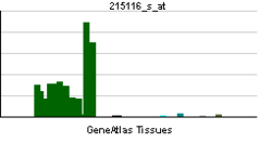
• receptor-mediated endocytosisSources: Amigo / QuickGO RNA expression pattern 
More reference expression data Orthologs Species Human Mouse Entrez 1759 13429 Ensembl ENSG00000106976 ENSMUSG00000026825 UniProt Q05193 Q6PDM5 RefSeq (mRNA) NM_001005336.1 NM_010065.2 RefSeq (protein) NP_001005336.1 NP_034195.2 Location (UCSC) Chr 9:
130.97 – 131.02 MbChr 2:
32.16 – 32.21 MbPubMed search [1] [2] Dynamin-1 is a protein that in humans is encoded by the DNM1 gene.[1][2]
This gene encodes a member of the dynamin subfamily of GTP-binding proteins. The encoded protein possesses unique mechanochemical properties used to tubulate and sever membranes, and is involved in clathrin-mediated endocytosis and other vesicular trafficking processes. Actin and other cytoskeletal proteins act as binding partners for the encoded protein, which can also self-assemble leading to stimulation of GTPase activity. More than sixty highly conserved copies of the 3' region of this gene are found elsewhere in the genome, particularly on chromosomes Y and 15. Alternatively spliced transcript variants encoding different isoforms have been described.[3]
Interactions
DNM1 has been shown to interact with Amphiphysin,[4][5][6][7][8] FNBP1,[9] NCK1,[10] PACSIN1,[9][11] Grb2[12][13] and SH3GL2.[4][14]
References
- ^ Obar RA, Collins CA, Hammarback JA, Shpetner HS, Vallee RB (Oct 1990). "Molecular cloning of the microtubule-associated mechanochemical enzyme dynamin reveals homology with a new family of GTP-binding proteins". Nature 347 (6290): 256–61. doi:10.1038/347256a0. PMID 2144893.
- ^ Newman-Smith ED, Shurland DL, van der Bliek AM (Jul 1997). "Assignment of the dynamin-1 gene (DNM1) to human chromosome 9q34 by fluorescence in situ hybridization and somatic cell hybrid analysis". Genomics 41 (2): 286–9. doi:10.1006/geno.1996.4596. PMID 9143509.
- ^ "Entrez Gene: DNM1 dynamin 1". http://www.ncbi.nlm.nih.gov/sites/entrez?Db=gene&Cmd=ShowDetailView&TermToSearch=1759.
- ^ a b Micheva, K D; Kay B K, McPherson P S (Oct. 1997). "Synaptojanin forms two separate complexes in the nerve terminal. Interactions with endophilin and amphiphysin". J. Biol. Chem. (UNITED STATES) 272 (43): 27239–45. doi:10.1074/jbc.272.43.27239. ISSN 0021-9258. PMID 9341169.
- ^ Wigge, P; Köhler K, Vallis Y, Doyle C A, Owen D, Hunt S P, McMahon H T (Oct. 1997). "Amphiphysin heterodimers: potential role in clathrin-mediated endocytosis". Mol. Biol. Cell (UNITED STATES) 8 (10): 2003–15. ISSN 1059-1524. PMC 25662. PMID 9348539. http://www.pubmedcentral.nih.gov/articlerender.fcgi?tool=pmcentrez&artid=25662.
- ^ McMahon, H T; Wigge P, Smith C (Aug. 1997). "Clathrin interacts specifically with amphiphysin and is displaced by dynamin". FEBS Lett. (NETHERLANDS) 413 (2): 319–22. doi:10.1016/S0014-5793(97)00928-9. ISSN 0014-5793. PMID 9280305.
- ^ Chen-Hwang, Mo-Chou; Chen Huey-Ru, Elzinga Marshall, Hwang Yu-Wen (May. 2002). "Dynamin is a minibrain kinase/dual specificity Yak1-related kinase 1A substrate". J. Biol. Chem. (United States) 277 (20): 17597–604. doi:10.1074/jbc.M111101200. ISSN 0021-9258. PMID 11877424.
- ^ Grabs, D; Slepnev V I, Songyang Z, David C, Lynch M, Cantley L C, De Camilli P (May. 1997). "The SH3 domain of amphiphysin binds the proline-rich domain of dynamin at a single site that defines a new SH3 binding consensus sequence". J. Biol. Chem. (UNITED STATES) 272 (20): 13419–25. doi:10.1074/jbc.272.20.13419. ISSN 0021-9258. PMID 9148966.
- ^ a b Kamioka, Yuji; Fukuhara Shigetomo, Sawa Hirofumi, Nagashima Kazuo, Masuda Michitaka, Matsuda Michiyuki, Mochizuki Naoki (Sep. 2004). "A novel dynamin-associating molecule, formin-binding protein 17, induces tubular membrane invaginations and participates in endocytosis". J. Biol. Chem. (United States) 279 (38): 40091–9. doi:10.1074/jbc.M404899200. ISSN 0021-9258. PMID 15252009.
- ^ Wunderlich, L; Faragó A, Buday L (Jan. 1999). "Characterization of interactions of Nck with Sos and dynamin". Cell. Signal. (ENGLAND) 11 (1): 25–9. doi:10.1016/S0898-6568(98)00027-8. ISSN 0898-6568. PMID 10206341.
- ^ Modregger, J; Ritter B, Witter B, Paulsson M, Plomann M (Dec. 2000). "All three PACSIN isoforms bind to endocytic proteins and inhibit endocytosis". J. Cell. Sci. (ENGLAND) 113 Pt 24: 4511–21. ISSN 0021-9533. PMID 11082044.
- ^ Miki, H; Miura K, Matuoka K, Nakata T, Hirokawa N, Orita S, Kaibuchi K, Takai Y, Takenawa T (Feb. 1994). "Association of Ash/Grb-2 with dynamin through the Src homology 3 domain". J. Biol. Chem. (UNITED STATES) 269 (8): 5489–92. ISSN 0021-9258. PMID 8119878.
- ^ Sastry, L; Cao T, King C R (Jan. 1997). "Multiple Grb2-protein complexes in human cancer cells". Int. J. Cancer (UNITED STATES) 70 (2): 208–13. doi:10.1002/(SICI)1097-0215(19970117)70:2<208::AID-IJC12>3.0.CO;2-E. ISSN 0020-7136. PMID 9009162.
- ^ Modregger, Jan; Schmidt Anne A, Ritter Brigitte, Huttner Wieland B, Plomann Markus (Feb. 2003). "Characterization of Endophilin B1b, a brain-specific membrane-associated lysophosphatidic acid acyl transferase with properties distinct from endophilin A1". J. Biol. Chem. (United States) 278 (6): 4160–7. doi:10.1074/jbc.M208568200. ISSN 0021-9258. PMID 12456676.
Further reading
- Sever S (2003). "Dynamin and endocytosis.". Curr. Opin. Cell Biol. 14 (4): 463–7. doi:10.1016/S0955-0674(02)00347-2. PMID 12383797.
- Wiejak J, Wyroba E (2003). "Dynamin: characteristics, mechanism of action and function.". Cell. Mol. Biol. Lett. 7 (4): 1073–80. PMID 12511974.
- Orth JD, McNiven MA (2003). "Dynamin at the actin-membrane interface.". Curr. Opin. Cell Biol. 15 (1): 31–9. doi:10.1016/S0955-0674(02)00010-8. PMID 12517701.
- Timm D, Salim K, Gout I, et al. (1995). "Crystal structure of the pleckstrin homology domain from dynamin.". Nat. Struct. Biol. 1 (11): 782–8. doi:10.1038/nsb1194-782. PMID 7634088.
- Downing AK, Driscoll PC, Gout I, et al. (1995). "Three-dimensional solution structure of the pleckstrin homology domain from dynamin.". Curr. Biol. 4 (10): 884–91. doi:10.1016/S0960-9822(00)00197-4. PMID 7850421.
- Ferguson KM, Lemmon MA, Schlessinger J, Sigler PB (1994). "Crystal structure at 2.2 A resolution of the pleckstrin homology domain from human dynamin.". Cell 79 (2): 199–209. doi:10.1016/0092-8674(94)90190-2. PMID 7954789.
- van der Bliek AM, Redelmeier TE, Damke H, et al. (1993). "Mutations in human dynamin block an intermediate stage in coated vesicle formation.". J. Cell Biol. 122 (3): 553–63. doi:10.1083/jcb.122.3.553. PMC 2119674. PMID 8101525. http://www.pubmedcentral.nih.gov/articlerender.fcgi?tool=pmcentrez&artid=2119674.
- Miki H, Miura K, Matuoka K, et al. (1994). "Association of Ash/Grb-2 with dynamin through the Src homology 3 domain.". J. Biol. Chem. 269 (8): 5489–92. PMID 8119878.
- Maruyama K, Sugano S (1994). "Oligo-capping: a simple method to replace the cap structure of eukaryotic mRNAs with oligoribonucleotides.". Gene 138 (1-2): 171–4. doi:10.1016/0378-1119(94)90802-8. PMID 8125298.
- Sontag JM, Fykse EM, Ushkaryov Y, et al. (1994). "Differential expression and regulation of multiple dynamins.". J. Biol. Chem. 269 (6): 4547–54. PMID 8308025.
- Grabs D, Slepnev VI, Songyang Z, et al. (1997). "The SH3 domain of amphiphysin binds the proline-rich domain of dynamin at a single site that defines a new SH3 binding consensus sequence.". J. Biol. Chem. 272 (20): 13419–25. doi:10.1074/jbc.272.20.13419. PMID 9148966.
- Ramjaun AR, Micheva KD, Bouchelet I, McPherson PS (1997). "Identification and characterization of a nerve terminal-enriched amphiphysin isoform.". J. Biol. Chem. 272 (26): 16700–6. doi:10.1074/jbc.272.26.16700. PMID 9195986.
- Ringstad N, Nemoto Y, De Camilli P (1997). "The SH3p4/Sh3p8/SH3p13 protein family: binding partners for synaptojanin and dynamin via a Grb2-like Src homology 3 domain.". Proc. Natl. Acad. Sci. U.S.A. 94 (16): 8569–74. doi:10.1073/pnas.94.16.8569. PMC 23017. PMID 9238017. http://www.pubmedcentral.nih.gov/articlerender.fcgi?tool=pmcentrez&artid=23017.
- McMahon HT, Wigge P, Smith C (1997). "Clathrin interacts specifically with amphiphysin and is displaced by dynamin.". FEBS Lett. 413 (2): 319–22. doi:10.1016/S0014-5793(97)00928-9. PMID 9280305.
- Wigge P, Köhler K, Vallis Y, et al. (1997). "Amphiphysin heterodimers: potential role in clathrin-mediated endocytosis.". Mol. Biol. Cell 8 (10): 2003–15. PMC 25662. PMID 9348539. http://www.pubmedcentral.nih.gov/articlerender.fcgi?tool=pmcentrez&artid=25662.
- Suzuki Y, Yoshitomo-Nakagawa K, Maruyama K, et al. (1997). "Construction and characterization of a full length-enriched and a 5'-end-enriched cDNA library.". Gene 200 (1-2): 149–56. doi:10.1016/S0378-1119(97)00411-3. PMID 9373149.
- Witke W, Podtelejnikov AV, Di Nardo A, et al. (1998). "In mouse brain profilin I and profilin II associate with regulators of the endocytic pathway and actin assembly.". EMBO J. 17 (4): 967–76. doi:10.1093/emboj/17.4.967. PMC 1170446. PMID 9463375. http://www.pubmedcentral.nih.gov/articlerender.fcgi?tool=pmcentrez&artid=1170446.
- Slepnev VI, Ochoa GC, Butler MH, et al. (1998). "Role of phosphorylation in regulation of the assembly of endocytic coat complexes.". Science 281 (5378): 821–4. doi:10.1126/science.281.5378.821. PMID 9694653.
PDB gallery 1dyn: CRYSTAL STRUCTURE AT 2.2 ANGSTROMS RESOLUTION OF THE PLECKSTRIN HOMOLOGY DOMAIN FROM HUMAN DYNAMIN2aka: Structure of the nucleotide-free myosin II motor domain from Dictyostelium discoideum fused to the GTPase domain of dynamin 1 from Rattus norvegicus2dyn: DYNAMIN (PLECKSTRIN HOMOLOGY DOMAIN) (DYNPH)Synaptic vesicle OtherCOPI COPII RME/Clathrin Caveolae Other/ungrouped Vesicle formationAdaptor protein complex 1: AP1AR · AP1B1 · AP1G1 · AP1G2 · AP1M1 · AP1M2 · AP1S1 · AP1S2 · AP1S3
Adaptor protein complex 2: AP2A1 · AP2A2 · AP2B1 · AP2M1 · AP2S1
Adaptor protein complex 3: AP3B1 · AP3B2 · AP3D1 · AP3M1 · AP3M2 · AP3S1 · AP3S2
Adaptor protein complex 4: AP4B1 · AP4E1 · AP4M1 · AP4S1
Coats: Retromer · TIP47Othersee also vesicular transport protein disorders
B memb: cead, trns (1A, 1C, 1F, 2A, 3A1, 3A2-3, 3D), othrCategories:- Human proteins
- Chromosome 9 gene stubs
Wikimedia Foundation. 2010.


