- Doppler fetal monitor
-
 Doppler fetal monitor
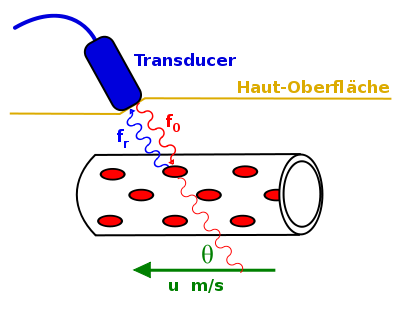
Invented in 1958 by Dr. Edward H. Hon[1] a Doppler fetal monitor or Doppler fetal heart rate monitor is a hand-held ultrasound transducer used to detect the heart beat of a fetus for prenatal care. It uses the Doppler effect to provide an audible simulation of the heart beat. Some models also display the heart rate in beats per minute. Use of this monitor is sometimes known as Doppler auscultation. Doppler fetal monitors are commonly referred to simply as "Dopplers".
Doppler fetal monitors provide information about the fetus similar that provided by a fetal stethoscope. One advantage of the Doppler fetal monitor over a (purely acoustic) fetal stethoscope is the electronic audio output, which allows people other than the user to hear the heartbeat. One disadvantage is the greater complexity and cost and the lower reliability of an electronic device.[citation needed]
Originally intended for use by health care professionals, this device is becoming popular for personal use.
Contents
Fetal heart rates
Starting at week 5 the fetal heart rate accelerates by 3.3 bpm per day for the next month.
The fetal heart begins to beat at approximately the same rate as the mother's, which is typically 80 to 85 bpm. The approximate fetal heart rate for weeks 5 to 9 (assuming a starting rate of 80):
- Week 5 starts at 80 and ends at 103 bpm
- Week 6 starts at 103 and ends at 126 bpm
- Week 7 starts at 126 and ends at 149 bpm
- Week 8 starts at 149 and ends at 172 bpm
- At week 9 the fetal heartbeat tends to beat within a range of 155 to 195 bpm.
At this point, the fetal heart rate begins to decrease, and generally falls within the range of 120 to 160 bpm by week 12.[2]
Types of Dopplers
Dopplers for home or hospital use differ in the following ways:
- Manufacturer: popular manufacturers are Nicolet, Huntleigh, Summit Doppler, EchoHeart, Ultrasound Technologies (Seward / Wakeling), Parks Medical Electronics (as Obstetrical Dopplers),and Sunray.
- Probe type: waterproof or not. Waterproof probes are used for water births.
- Frequency: 2- or 3-MHz probes. Most practitioners can find the heart rate with either probe. A 3-MHz probe is recommended to detect a heart rate in early pregnancy (8–10 weeks gestation). A 2-MHz probe is recommended for pregnant women who are overweight. A new 5-MHz EchoHeart Transvaginal Fetal Doppler Probe aids in the detection of fetal heart tones (FHT) early in pregnancy (6–8 weeks) and for patients who have a retroverted uterus or throughout pregnancy for FHT detection for women who are obese.
- Heart rate display: some Dopplers automatically display the heart rate; for others the fetal heart rate must be counted and timed by the practitioner.
The generic use of the word "Sonicaid" for Doppler fetal monitors comes from the products of the UK company Sonicaid Ltd. Sonicaid products included the D205/206 portable fetal Dopplers and FM2/3/4 series of fetal monitors. The company was acquired by Oxford Instruments in 1987 to form Oxford Sonicaid.
See also
- Ultrasonic sensor
- Ultrasound
- Cardiotocograph
- Nonstress test
- Auscultation
References
- ^ Roger K. Freeman, Thomas J. Garite, Michael P. Nageotte, Fetal Heart Rate Monitoring Third Edition, 2003, p. 3. "The earliest preliminary report of FHR monitoring came in 1958 from Edward Hon, MD,... via fetal ECG monitor on the maternal abdomen." Google Books citation
- ^ FetalSure. Fetal Heart and Heartbeat Facts. Available at http://www.fetalsure.com/fetal-heart.html. Retrieved 9 August 2010.
Categories:


Wikimedia Foundation. 2010.