- Silver stain
-
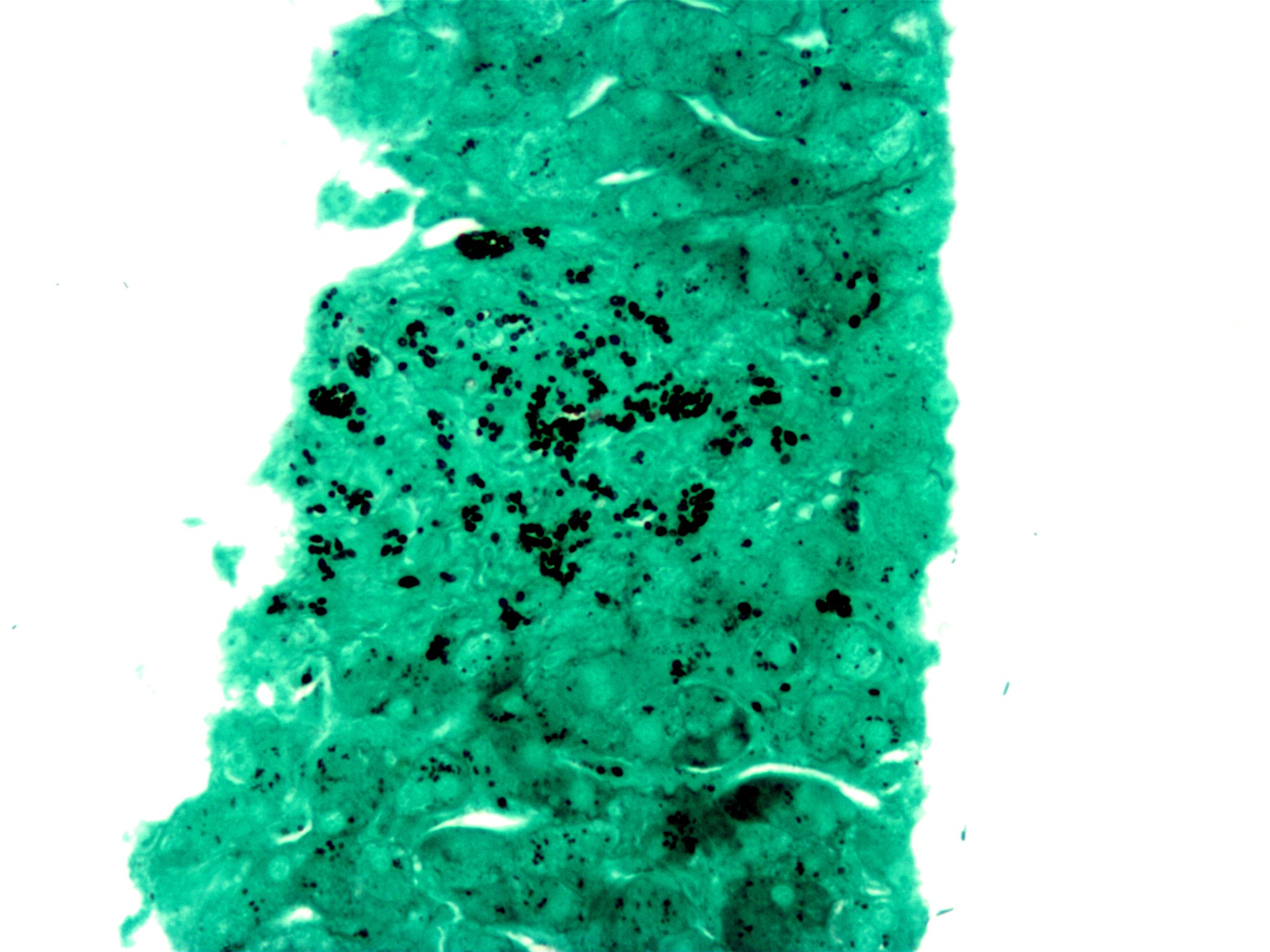
 A silver stain (GMS) demonstrating the fungus histoplasma (black round balls) in a liver biopsy.
Silver staining is the use of silver to selectively alter the appearance of the target.
Contents
Use in medicine
It is used to stain histologic sections. This kind of staining is important especially to show proteins (for example type III collagen) and DNA. It is used to show both substances inside and outside cells. Silver staining is also used in temperature gradient gel electrophoresis and in polyacrylamide gels.
 A number of DNA samples from specimens of Littorina plena amplified using polymerase chain reaction with primers targeting a variable simple sequence repeat (SSR, a.k.a. microsatellite) locus. Samples have been run on a 5% polyacrylamide gel and visualized using silver staining.
Some cells are argentaffin. These reduce silver solution to metallic silver after formalin fixation. Other cells are argyrophilic. These reduce silver solution to metallic silver after being exposed to the stain that contains a reductant, for example hydroquinone or formalin.
Silver nitrate forms insoluble silver phosphate with phosphate ions; this method is known as the Von Kossa Stain. When subjected to a reducing agent, usually hydroquinone, it forms black elementary silver. This is used for study of formation of calcium phosphate particles during bone growth.
Silver staining is used in light microscopy. The metallic silver particles are deposited on sensitised reticulin fibres and are then easily seen in the microscopic preparations.
Silver stain aids in the perception of reticular fibers.[1]
Karyotype analysis
Silver staining is also used in karyotype analysis. Silver nitrate stains the nucleolar organization region-associated protein. This yields a dark region where the silver is deposited, denoting the activity of rRNA genes within the NOR. Chromosomes 13, 14, 15, 21, and 22 have NORs.
Organisms highlighted
Pseudomonas,[2] Legionella, Leptospira, H. pylori, and fungi like Pneumocystis and Candida.
Methenamine silver stains
There are several silver stains incorporating methenamine, including:
- Grocott's methenamine silver stain, used widely as a screen for fungal organisms.
- Jones' stain, a methenamine silver-Periodic acid-Schiff that stains for basement membrane, availing to view the "spiked" GBM associated with membranous glomerulonephritis.
Silver staining of SDS Polyacrylamide gels
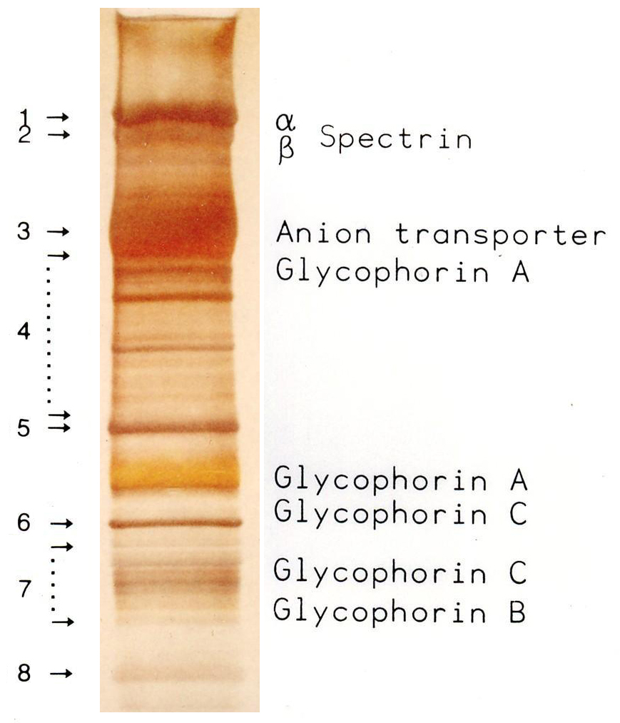
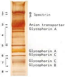 Red blood cell membrane proteins separated by SDS-Page and silverstained [3]
Camillo Golgi perfected the silver staining for the study of the nervous system. Golgi's method stains a limited number of cells at random in their entirety.[4] The exact chemical mechanism by which this happens is still largely unknown.[5] Silver staining was introduced by Kerenyi and Gallyas as a sensitive procedure to detect trace amounts of proteins in gels.[6] The technique has been extended to the study of other biological macromolecules that have been separated in a variety of supports.[7] Classical Coomassie Brilliant Blue staining can usually detect a 50 ng protein band, Silver staining increases the sensitivity typically 50 times. Many variables can influence the colour intensity and every protein has its own staining characteristics; clean glassware, pure reagents and water of highest purity are the key points to successful staining.[8]
Use in art
Silver staining is also a technique in traditional stained glass to produce the yellow, brown or amber shading when painting on glass. It is a technique that is often used for realistic hair colors. It was discovered in the 14th Century but was not originally used very frequently.
References
- ^ Schwint OA, Labraga M, Cervino CO, Haffar M, Sequeiros PH, Marcos HJ (2004). "A modification of the staining technique of reticular fibres for image analysis of the cardiac collagen network". Cardiovasc. Pathol. 13 (4): 213–20. doi:10.1016/S1054-8807(03)00153-4. PMID 15210137. http://linkinghub.elsevier.com/retrieve/pii/S1054880703001534.
- ^ Barnini S, Dodi C, Campa M (2004). "Enhanced resolution of random amplified polymorphic DNA genotyping of Pseudomonas aeruginosa". Lett. Appl. Microbiol. 39 (3): 274–7. doi:10.1111/j.1472-765X.2004.01576.x. PMID 15287874. http://www3.interscience.wiley.com/resolve/openurl?genre=article&sid=nlm:pubmed&issn=0266-8254&date=2004&volume=39&issue=3&spage=274.
- ^ Hempelmann E, Götze O (1984). "Characterization of membrane proteins by polychromatic silver staining". Hoppe Seyler's Z Physiol Chem 365: 241–242.
- ^ Grant G (Oct 2007). "How the 1906 Nobel Prize in Physiology or Medicine was shared between Golgi and Cajal". Brain Res Rev 55 (2): 490–498. doi:10.1016/j.brainresrev.2006.11.004. PMID 17306375.
- ^ Golgi C (1873). "Sulla struttura della sostanza grigia del cervello.". Gazzetta Medica Italiana (Lombardia) 33: 244–246.
- ^ Kerenyi L, Gallyas F (1973). "Über Probleme der quantitiven Auswertung der mit physikalischer Entwicklung versilberten Agarelektrophoretogramme". Clin. Chim. Acta 47 (3): 425–436. doi:10.1016/0009-8981(73)90276-3. PMID 4744834.
- ^ Switzer RC 3rd, Merril CR, Shifrin S (Sep 1979). "A highly sensitive silver stain for detecting proteins and peptides in polyacrylamide gels.". Anal Biochem. 98 (1): 231–237. doi:10.1016/0003-2697(79)90732-2. PMID 94518.
- ^ Hempelmann E, Schulze M, Götze O (1984). "Free SH-groups are important for the polychromatic staining of proteins with silver nitrat". Neuhof V (ed)Electrophoresis '84, Verlag Chemie Weinheim 1984: 328–330.
External links
- MedEd at Loyola Histo/practical/stains/hp2-55.html
- [1] Hempelmann E. SDS-Protein PAGE and Proteindetection by Silverstaining and Immunoblotting of Plasmodium falciparum proteins. in: Moll K, Ljungström J, Perlmann H, Scherf A, Wahlgren M (eds) Methods in Malaria Research, 5th edition, 2008, 263-266
Stains Iron/Hemosiderin Lipids Carbohydrates Amyloid Bacteria Gram staining (Methyl violet/Gentian violet, Safranin) · Ziehl–Neelsen stain/acid-fast (Carbol fuchsin/Fuchsine, Methylene blue) · Auramine-rhodamine stain (Auramine O, Rhodamine B)Connective tissue Other H&E stain (Haematoxylin, Eosin Y) · Silver stain (Grocott's methenamine silver stain, Warthin–Starry stain) · Methyl blue · Wright's stain · Giemsa stain · Gömöri trichrome stain · Neutral red · Janus Green BTissue stainability Categories:- Staining
- Electron microscopy stains
Wikimedia Foundation. 2010.