- Eumycetoma
-
Mycetoma Classification and external resources 
Madura FootICD-10 B47.0 DiseasesDB 8472 eMedicine med/30 derm/280 derm/147 MeSH D008271 - "Mycetoma" redirects here. For the bacterial disease formerly known as "actinomycetoma", see Actinomycosis.
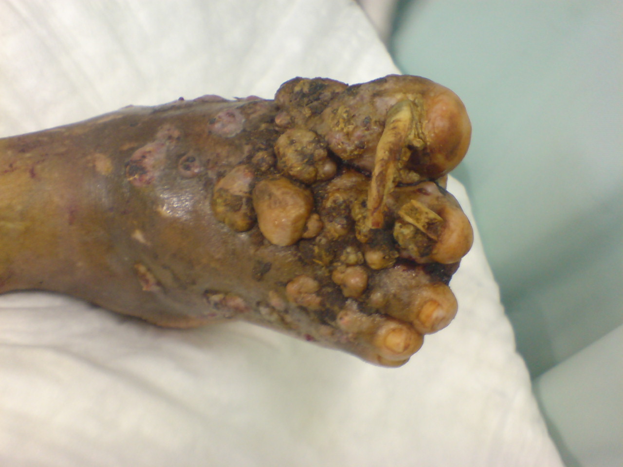
Eumycetoma is a chronic, specific, granulomatous,[1] fungal disease.[2] It mainly affects the foot; and Mycetoma pedis is also known as Madura foot. This infection is endemic in Africa, India, and Central and South America.[3]
Contents
Causes and presentation
Eumycetoma usually involves the subcutaneous tissue after a traumatic inoculation of the causative organism. Tumefaction and formation of sinus tracts characterize mycetoma. The sinuses usually discharge purulent and seropurulent exudate containing grains. It may spread to involve the skin and the deep structures resulting in destruction, deformity and loss of function; very occasionally it could be fatal.
Epidemiology
The true incidence and the geographical distribution of mycetoma throughout the world is not exactly known due to the nature of the disease which is usually painless, slowly progressive which may lead to the late presentation of the majority of patients. Mycetoma has a worldwide distribution but this is extremely uneven. It is endemic in tropical and subtropical regions. The African continent seems to have the highest prevalence. It is found in what is known as the mycetoma belt stretching between the latitudes of 15 south and 30 north. The belt includes Sudan, Somalia, Senegal, India, Yemen, Mexico, Venezuela, Colombia, Argentina and others.
Mycetoma infection, Madura foot or maduromycosis, was originally described in Sanskrit in the Vedic texts from India. The first English language accounts occurred much later in the area of Madras (aka Chennai).
The geographical distribution of the individual mycetoma organism shows considerable variations, which can be convincingly explained on an environmental basis. Areas where mycetoma prevails are relatively arid zones with a short rainy season with a relative humidity.
The organisms are usually present in the soil in the form of grains. The infecting agent is implanted into the host tissue through a breach in the skin produced by trauma caused by sharp objects such as thorn pricks, stone or splinters.
Pathogenesis
The disease is usually acquired while performing agricultural work, and it generally afflicts men between 20 and 40 years old. The disease is acquired by contacting grains of fungal spores that have been discharged onto the soil. Infection usually involves an open area or break in the skin. Pseudoallescheria boydii is one of many fungi spp. that causes the fungal form of madura foot (see below). The disease is characterized by a yogurt-like discharge upon maturation of the infection. Hematogenous or lymphatic spread is uncommon. Infections normally start in the foot or hand and travel up the leg or arm.
Eumycetoma may be one of several varieties, depending upon color of the granulous discharge:
- red
- Actinomadura pelletieri
- white or yellow
- Acremonium strictum
- Actinomadura madurae
- Aspergillus nidulans
- Noetestudina rosatii
- Phaeoacremonium krajdenii[4]
- Pseudallescheria boydii[5]
- black
- Curvularia lunata
- Exophiala jeanselmei[6]
- Leptosphaeria senegalensis
- Leptosphaeria tompkinsii
- Madurella grisea[7]
- Madurella mycetomatis[8]
- Pyrenochaeta romeroi
The bacterial mycetoma species Nocardia (including Nocardia asteroides and Nocardia brasiliensis) produces yellow discharge, and Streptomyces (including Streptomcyes somaliensis) produce yellow or red discharge. These are not causes of Eumycetoma.
The further course of the sinuses differs according to the mycetoma present. In the black variety, the infection spreads mainly subcutaneously. In the red and yellow varieties deep spread occurs early, and muscle and underlying bones become infiltrated, but unexpectedly, nerves and tendons are highly resistant to invasion.[3]
Diagnosis
Diagnosis of mycetoma is usually accomplished by radiology, ultrasound or by fine needle aspiration of the fluid within an afflicted area of the body. It depends upon isolating the causative organism along with a knowledge of local endemic infection.
 Madura Foot X-Ray
Differential diagnosis
Following is the differential diagnosis:
- Tuberculous ulcer
- Kaposi's sarcoma
- Tropical ulcer[3]
Treatment
There are several clinical treatments available for this disease. They include surgery, ketoconazole,[9] voriconazole,[10] itraconazole and amputation. There is no vaccine for mycetoma.
Scientists at such institutions as The Mycetoma Research Center at The University of Khartoum in the Sudan are working on a cure.
References
- ^ Motswaledi HM, Mathekga K, Sein PP, Nemutavhanani DL (August 2009). "Paecilomyces lilacinus eumycetoma". Int. J. Dermatol. 48 (8): 858–61. doi:10.1111/j.1365-4632.2008.04047.x. PMID 19659864. http://www3.interscience.wiley.com/resolve/openurl?genre=article&sid=nlm:pubmed&issn=0011-9059&date=2009&volume=48&issue=8&spage=858.
- ^ Brownell I, Pomeranz M, Ma L (2005). "Eumycetoma". Dermatol. Online J. 11 (4): 10. PMID 16403382. http://dermatology.cdlib.org/114/NYU/NYUtexts/0419058.html.
- ^ a b c Hamilton Bailey's Demonstrations of Physical Signs in Clinical Surgery ISBN 0-7506-0625-8
- ^ Hemashettar BM, Siddaramappa B, Munjunathaswamy BS, et al. (December 2006). "Phaeoacremonium krajdenii, a cause of white grain eumycetoma". J. Clin. Microbiol. 44 (12): 4619–22. doi:10.1128/JCM.01019-06. PMC 1698411. PMID 17005754. http://jcm.asm.org/cgi/pmidlookup?view=long&pmid=17005754.
- ^ "Filamentous Fungi". http://pathmicro.med.sc.edu/mycology/mycology-5.htm.
- ^ Severo LC, Oliveira FM, Vettorato G, Londero AT (March 1999). "Mycetoma caused by Exophiala jeanselmei. Report of a case successfully treated with itraconazole and review of the literature". Rev Iberoam Micol 16 (1): 57–9. PMID 18473595. http://www.reviberoammicol.com/pubmed_linkout.php?16p57.
- ^ Vilela R, Duarte OM, Rosa CA, et al. (November 2004). "A case of eumycetoma due to Madurella grisea in northern Brazil". Mycopathologia 158 (4): 415–8. doi:10.1007/s11046-004-2844-y. PMID 15630550. http://www.kluweronline.com/art.pdf?issn=0301-486X&volume=158&page=415.
- ^ Ahmed AO, Desplaces N, Leonard P, et al. (December 2003). "Molecular detection and identification of agents of eumycetoma: detailed report of two cases". J. Clin. Microbiol. 41 (12): 5813–6. doi:10.1128/JCM.41.12.5813-5816.2003. PMC 309011. PMID 14662990. http://jcm.asm.org/cgi/pmidlookup?view=long&pmid=14662990.
- ^ Capoor MR, Khanna G, Nair D, et al. (April 2007). "Eumycetoma pedis due to Exophiala jeanselmei". Indian J Med Microbiol 25 (2): 155–7. doi:10.4103/0255-0857.32726. PMID 17582190. http://www.ijmm.org/article.asp?issn=0255-0857;year=2007;volume=25;issue=2;spage=155;epage=157;aulast=Capoor.
- ^ Loulergue P, Hot A, Dannaoui E, et al. (December 2006). "Successful treatment of black-grain mycetoma with voriconazole". Am. J. Trop. Med. Hyg. 75 (6): 1106–7. PMID 17172376. http://www.ajtmh.org/cgi/pmidlookup?view=long&pmid=17172376.
External links
- Centraalbureau voor Schimmelcultures/The Fungus Research Center Fungal Biodiversity Center
- DermNet fungal/mycetoma
- DermAtlas 336868008
Infectious diseases · Mycoses and Mesomycetozoea (B35–B49, 110–118) Superficial and
cutaneous
(dermatomycosis):
Tinea=skin;
Piedra (exothrix/
endothrix)=hairBy locationTinea barbae/Tinea capitis (Kerion) · Tinea corporis (Ringworm, Dermatophytid) · Tinea cruris · Tinea manuum · Tinea pedis (Athlete's foot) · Tinea unguium/Onychomycosis (White superficial onychomycosis · Distal subungual onychomycosis · Proximal subungual onychomycosis)
Tinea corporis gladiatorum · Tinea faciei · Tinea imbricata · Tinea incognito · FavusBy organismEpidermophyton floccosum · Microsporum canis · Microsporum audouinii · Trichophyton interdigitale/mentagrophytes · Trichophyton tonsurans · Trichophyton schoenleini · Trichophyton rubrumOtherHortaea werneckii (Tinea nigra) · Piedraia hortae (Black piedra)Subcutaneous,
systemic,
and opportunisticDimorphic
(yeast+mold)Coccidioides immitis/Coccidioides posadasii (Coccidioidomycosis, Disseminated coccidioidomycosis, Primary cutaneous coccidioidomycosis. Primary pulmonary coccidioidomycosis) · Histoplasma capsulatum (Histoplasmosis, Primary cutaneous histoplasmosis, Primary pulmonary histoplasmosis, Progressive disseminated histoplasmosis) · Histoplasma duboisii (African histoplasmosis) · Lacazia loboi (Lobomycosis) · Paracoccidioides brasiliensis (Paracoccidioidomycosis)OtherBlastomyces dermatitidis (Blastomycosis, North American blastomycosis, South American blastomycosis) · Sporothrix schenckii (Sporotrichosis) · Penicillium marneffei (Penicilliosis)Yeast-likeCandida albicans (Candidiasis, Oral, Esophageal, Vulvovaginal, Chronic mucocutaneous, Antibiotic candidiasis, Candidal intertrigo, Candidal onychomycosis, Candidal paronychia, Candidid, Diaper candidiasis, Congenital cutaneous candidiasis, Perianal candidiasis, Systemic candidiasis, Erosio interdigitalis blastomycetica) · C. glabrata · C. tropicalis · C. lusitaniae · Pneumocystis jirovecii (Pneumocystosis, Pneumocystis pneumonia)Mold-likeAspergillus (Aspergillosis, Aspergilloma, Allergic bronchopulmonary aspergillosis, Primary cutaneous aspergillosis) · Exophiala jeanselmei (Eumycetoma) · Fonsecaea pedrosoi/Fonsecaea compacta/Phialophora verrucosa (Chromoblastomycosis) · Geotrichum candidum (Geotrichosis) · Pseudallescheria boydii (Allescheriasis)Entomophthorales
(Entomophthoramycosis)Basidiobolus ranarum (Basidiobolomycosis) · Conidiobolus coronatus/Conidiobolus incongruus (Conidiobolomycosis)Enterocytozoon bieneusi/Encephalitozoon intestinalisMesomycetozoea Ungrouped Alternariosis · Fungal folliculitis · Fusarium (Fusariosis) · Granuloma gluteale infantum · Hyalohyphomycosis · Otomycosis · PhaeohyphomycosisCategories:- Mycosis-related cutaneous conditions

Wikimedia Foundation. 2010.