- Extraocular muscles
-
Extraocular muscles 
MRI scan showing lateral and medial rectus muscles. Latin musculi externi bulbi oculi Origin Insertion Artery Nerve CNIII, IV, VI Actions The extraocular muscles are the six muscles that control the movements of the (human) eye (there are four in bovines). The actions of the extraocular muscles depend on the position of the eye at the time of muscle contraction.
Contents
List of muscles
Muscle Innervation Origin Insertion Primary function Secondary function Tertiary function Inserted into the sclera distance Superior rectus Superior branch of oculomotor nerve Annulus of Zinn from tendinous ring eye (anterior, superior surface) Elevation Intorsion Adduction[1] 7.9mm Inferior rectus Inferior branch of oculomotor nerve Annulus of Zinn from tendinous ring eye (anterior, inferior surface) Depression Extorsion Adduction[1] 6.6mm Lateral rectus Abducens nerve Annulus of Zinn from tendinous ring eye (anterior, lateral surface) Abduction 7.0mm Medial rectus Inferior branch of oculomotor nerve Annulus of Zinn from tendinous ring eye (anterior, medial surface) Adduction 5.8mm Superior oblique Trochlear nerve Annulus of Zinn via the Trochlea of superior oblique which forms a 'pulley system'. eye (posterior, superior, lateral surface) Intorsion Depression Abduction[1] Inferior oblique Inferior branch of oculomotor nerve Maxillary bone eye (posterior, inferior, lateral surface) Extorsion Elevation Abduction[1] Importance
 Extraocular muscles
Since only the fovea provides sharp distinct vision, the eye must move to follow a target. It must be precise and fast. This is seen in scenarios like reading, wherein the reader must shift gaze constantly, or following a small object like a golf ball, in which the extraocular muscles must lead the eye to follow the head movements. Although under voluntary control, most movement is done without thinking, such as those based on head or other body movement, or movement of objects in the area. Researchers still have some work in order to find the parallel nature of the environment-based (involuntary) and voluntary control.[2]
Innervation
The nuclei or bodies of these nerves are found in the brain stem. The nuclei of the abducens and oculomotor nerves are connected. This is important in coordinating motion of the lateral rectus in one eye and the medial action on the other. In one eye, in two antagonistic muscles, like the lateral and medial recti, contraction of one leads to inhibition of the other. Muscles shows small degrees of activity even when resting, keeping the muscles taut. This "tonic" activity is brought on by discharges of the motor nerve to the muscle.[2]
Actions[3]
Note that intorsion and extorsion are not included in the following table; their actions are accounted for via summation of other actions.
Medial (towards nose) Lateral (towards temple) Elevation, abduction:
Inferior obliqueElevation, adduction:
Superior rectusAdduction:
Medial rectusAbduction:
Lateral rectusDepression, abduction:
Superior obliqueDepression, adduction:
Inferior rectus- These motions are only for eye examinations. Note that they are different from the intrinsic motor functions of each muscle. This is done in an exam to separate out the muscle being tested specifically.
In an eye examination, the inability of the patient to move the eye in the specified direction can indicate a problem with the associated muscle, and the nerve associated with that muscle.
- Coordination of Movement Between Both Eyes
Intermediate directions are controlled by simultaneous actions of multiple muscles. When one shifts the gaze horizontally, one eye will move laterally (toward the side) and the other will move medially (toward the midline). This may be neurally coordinated by the central nervous system, to make the eyes move together and almost involuntarily. This is a key factor in the study of squint, namely, the inability of the eyes to be directed to one point.
There are two main kinds of movement: conjugate movement (the eyes move in the same direction) and disjunctive (opposite directions). The former is typical when shifting gaze right or left, the latter is convergence of the two eyes on a near object. Disjunction can be performed voluntarily, but is usually triggered by the nearness of the target object. A "see-saw" movement, namely, one eye looking up and the other down, is possible, but not voluntarily; this effect is brought on by putting a prism in front of one eye, so the relevant image is apparently displaced. To avoid double vision from non-corresponding points, the eye with the prism must move up or down, following the image passing through the prism. Likewise conjugate torsion (rolling) on the anteroposterior axis (from the front to the back) can occur naturally, such as when one tips one's head to one shoulder; the torsion, in the opposite direction, keeps the image vertical.
The muscles show little inertia - a shutdown of one muscle is not due to checking of the antagonist, so the motion is not ballistic.[2]
Paths
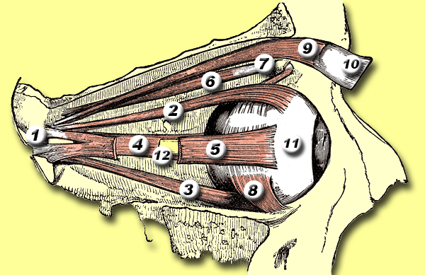
 View of right eye from the right:
1 = Annulus tendineus communis
2 = Superior rectus muscle
3 = Inferior rectus muscle
4 = Medial rectus muscle
5 = Lateral rectus muscle
6 = Superior oblique muscle
7 = Trochlea of superior oblique
8 = Inferior oblique muscle
9 = Levator palpebrae superioris muscle
10 = Eyelid
11 = Eyeball
12 = Optic nerveFive with paths from annulus of zinn
Five of the extraocular muscles have their origin in the back of the orbit in a fibrous ring called the annulus of Zinn.
Four of these then course forward through the orbit and insert onto the globe on its anterior half (i.e., in front of the eye's equator). These muscles are named after their straight paths, and are called the four rectus muscles, or four recti.[2]
- superior rectus - inserts on the globe at 2
- inferior rectus - inserts on the globe at 3
- medial rectus - inserts on the globe at 4
- lateral rectus - inserts on the globe at 5
(Note that lateral and medial are relative to the subject, with lateral toward the side and medial toward the midline, thus the medial rectus is the muscle closest to the nose).
Two with more complex paths
The other two extraocular muscles follow more complicated paths.
- The superior oblique muscle originates at the back of the orbit (a little closer to the medial rectus, though medial to it, getting rounder as it [2] courses forward to a rigid, cartilaginous pulley, called the trochlea, on the upper, nasal wall of the orbit. The muscle becomes tendinous about 10mm before it passes through the pulley, turning sharply across the orbit, and inserts on the lateral, posterior part of the globe. Thus, the superior oblique travels posteriorly for the last part of its path, going over the top of the eye. Due to its unique path, the superior oblique, when activated, pulls the eye downward and medially.[4]
- The last muscle is the inferior oblique, which originates at the lower front of the nasal orbital wall, and passes under the LR to insert on the lateral, posterior part of the globe. Thus, the inferior oblique pulls the eye upward and laterally .[4][5][6]
Rolling
The superior and inferior recti are not strictly vertical. The oblique pull of the obliques causes a rolling opposite each other. Although bearing mutual strict antagonism, the superior and inferior rectus team up with the inferior and superior oblique to move the eye up or down, respectively. The extent of rolling in the recti is less than the oblique, and opposite from it.[2]
Mnemonics
A good mnemonic to remember which muscles are innervated by what nerve is to paraphrase it as a molecular equation: LR6SO4R3.[7] or (LR6SO4)3 i.e. "LR 6 SO 4 Whole 3."
- Lateral Rectus - Cranial Nerve VI
- Superior Oblique - Cranial Nerve IV
- the Rest of the muscles - Cranial Nerve III.
Another way to remember which nerves innervate which muscles is to understand the meaning behind all of the Latin words.
- The fourth cranial nerve, the trochlear, is so named because the muscle it innervates, the superior oblique, runs through a little fascial pulley that changes its direction of pull (the trochlea of superior oblique). This pulley exists in the superiomedial corner of each orbit, and "trochl-" is Latin for "pulley."
- The sixth cranial nerve, the abducens, is so named because it controls the lateral rectus, which abducts the eye (rotates it laterally) upon contraction.
- The third cranial nerve, the oculomotor, is so named because it is in charge of the movement (motor) of the eye (oculo-). It controls all of the other muscles.
See also
Additional images
-

Nerves of the orbit. Seen from above.
-

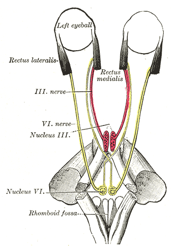
Figure showing the mode of innervation of the Recti medialis and lateralis of the eye.
-

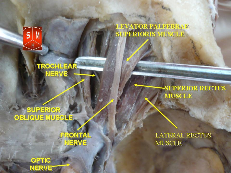
Dissection showing origins of right ocular muscles, and nerves entering by the superior orbital fissure.
References
- ^ a b c d http://www.tedmontgomery.com/the_eye/eom.html
- ^ a b c d e f "eye, human."Encyclopædia Britannica from Encyclopædia Britannica 2006 Ultimate Reference Suite DVD 2009
- ^ http://www.anat.stonybrook.edu/HBA531/EGA/EGA_2003_8.pdf
- ^ a b A case of mistaken muscles - Ahmed and Ali 324 (7343): 962 - BMJ
- ^ Roger H.S. Carpenter (1988); Movements of the Eyes (2nd ed.). Pion Ltd, London. ISBN 0-85086-109-8.
- ^ Westheimer Gerald, McKee Suzanne P (1975); "Visual acuity in the presence of retinal-image motion". Journal of the Optical Society of America 65(7), 847-50.
- ^ {{Medicalmnemonics.com|572|||}}
External links
- neuro/637 at eMedicine - "Extraocular Muscles, Actions"
- Atlas of anatomy at UMich eye_13
List of muscles of head and neck: the head (TA A04.1, GA 4.378) Extraocular (CN III, IV, VI) oblique (inferior, superior) · rectus (superior, inferior, medial, lateral) · levator palpebrae superioris (superior tarsal)Mastication (CN V3) masseter · temporalis (sphenomandibularis) · pterygoid (lateral, medial)
fascia: Masseteric fascia · Temporal fascia · Deep portion: cementomaxillary tendon · Superficial portion: cementomandibular tendonFacial (CN VII) levator anguli oris · levator labii superioris · zygomaticus (major, minor)
orbicularis oris · risorius · buccinator
depressor anguli oris · depressor labii inferioris · mentalisPalate/fauces (CN IX, X, XI)
(except TVP=V3)veli palatini (tensor, levator) · musculus uvulae · palatopharyngeus (to pharynx) · palatoglossus (to tongue)Tongue (CN XII) extrinsic (genioglossus, hyoglossus/chondroglossus, styloglossus, and palatoglossus) · intrinsic (superior longitudinal, inferior longitudinal, transverse, vertical)Muscles of orbit / Extraocular muscles (TA A15.2.07, GA 10.1021) Eyelid Globe Head and neck anatomy – accessory visual structures (TA 15.2.7, TH H3.11.08.6, GA 10.1021) Eyelid Tarsus (Meibomian pelicle) • Medial palpebral ligament • Epicanthic fold • Meibomian gland • Ciliary glands • Eyelash
Gland of ZeisLacrimal apparatus Lacrimal lake • Lacrimal gland • Lacrimal canaliculi • Lacrimal punctum • Lacrimal papilla • Nasolacrimal duct • Lacrimal sac • Lacrimal caruncle • Krause's glandsOther Periorbita • Orbital septum • Tenon's capsule • Suspensory ligament of eyeball
Conjunctiva (Plica semilunaris)
Extraocular muscles (Trochlea of superior oblique)M: EYE
anat(g/a/p)/phys/devp/prot
noco/cong/tumr, epon
proc, drug(S1A/1E/1F/1L)
Categories:- Muscles of the head and neck
- Eye anatomy


Wikimedia Foundation. 2010.