- ImageJ
-
ImageJ 
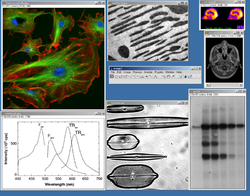
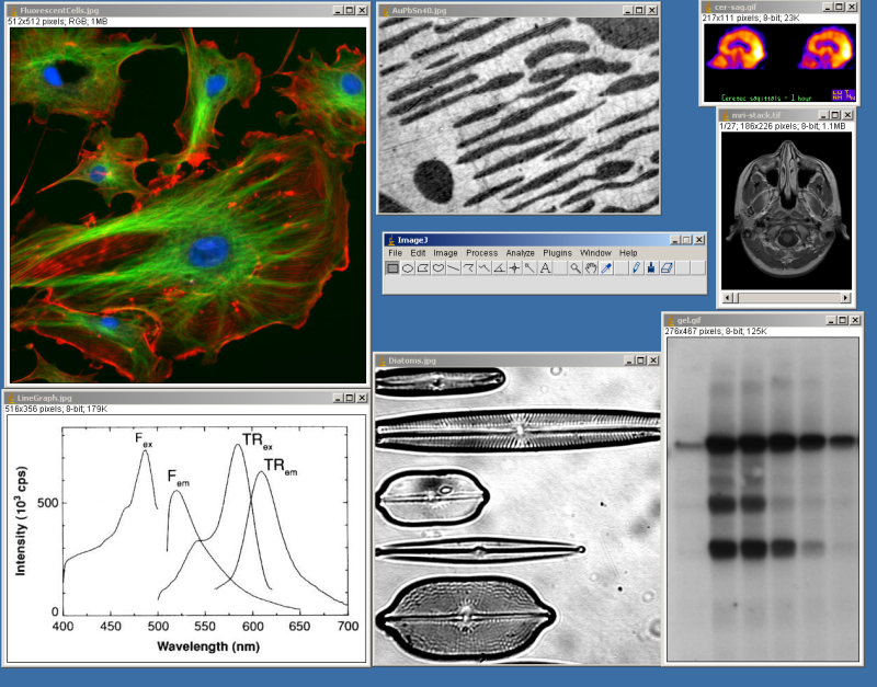
Screenshot of ImageJDeveloper(s) Wayne Rasband (NIH) Stable release 1.45s / October 29, 2011 Operating system Any (Java-based) Type Image processing License Public Domain Website http://rsb.info.nih.gov/ij/ ImageJ is a public domain, Java-based image processing program developed at the National Institutes of Health.[1] ImageJ was designed with an open architecture that provides extensibility via Java plugins and recordable macros.[2] Custom acquisition, analysis and processing plugins can be developed using ImageJ's built-in editor and a Java compiler. User-written plugins make it possible to solve many image processing and analysis problems, from three-dimensional live-cell imaging,[3] to radiological image processing,[4] multiple imaging system data comparisons[5] to automated hematology systems.[6] ImageJ's plugin architecture and built in development environment has made it a popular platform for teaching image processing.[7][8]
ImageJ can be run as an online applet, a downloadable application, or on any computer with a Java 5 or later virtual machine. Downloadable distributions are available for Microsoft Windows, Mac OS, Mac OS X, Linux, and the Sharp Zaurus PDA. The source code for ImageJ is freely available.[9]
The project developer, Wayne Rasband, is at the Research Services Branch of the National Institute of Mental Health.
Contents
Features
ImageJ can display, edit, analyze, process, save, and print 8-bit, 16-bit and 32-bit images. It can read many image formats including TIFF, PNG, GIF, JPEG, BMP, DICOM, FITS, as well as raw formats. ImageJ supports image stacks, a series of images that share a single window, and it is multithreaded, so time-consuming operations can be performed in parallel on multi-CPU hardware. ImageJ can calculate area and pixel value statistics of user-defined selections and intensity thresholded objects. It can measure distances and angles. It can create density histograms and line profile plots. It supports standard image processing functions such as logical and arithmetical operations between images, contrast manipulation, convolution, Fourier analysis, sharpening, smoothing, edge detection and median filtering. It does geometric transformations such as scaling, rotation and flips. The program supports any number of images simultaneously, limited only by available memory.
History
Prior to the release of ImageJ in 1997, a similar freeware image analysis program known as NIH Image had been developed for Macintosh computers running pre-Mac OS X operating systems. Further development of this code continues in the form of Image SXM, a variant tailored for physical research of scanning microscope images. A Windows version – ported by Scion Corporation, so called Scion Image for Windows – was also developed. Both versions are still available.[10]
See also
- Microscope image processing
- Fiji (Fiji Is Just ImageJ), an image processing package based on ImageJ
- Rapidminer Image Processing Extension - tool for image processing and image mining
- Bitplane - producers of image processing software with ImageJ compatibility
- CoLocalizer Pro - software application for quantification of colocalization in fluorescence microscopy images
References
- ^ Collins TJ (July 2007). "ImageJ for microscopy". BioTechniques 43 (1 Suppl): 25–30. doi:10.2144/000112517. PMID 17936939.
- ^ Girish V, Vijayalakshmi A (2004). "Affordable image analysis using NIH Image/ImageJ". Indian J Cancer 41 (1): 47. PMID 15105580. http://www.bioline.org.br/request?cn04009.
- ^ Eliceiri K, Rueden C (2005). "Tools for visualizing multidimensional images from living specimens". Photochem Photobiol 81 (5): 1116–22. doi:10.1562/2004-11-22-IR-377. PMID 15807634.
- ^ Barboriak D, Padua A, York G, Macfall J (2005). "Creation of DICOM—Aware Applications Using ImageJ". J Digit Imaging 18 (2): 91–9. doi:10.1007/s10278-004-1879-4. PMC 3046706. PMID 15827831. http://www.pubmedcentral.nih.gov/articlerender.fcgi?tool=pmcentrez&artid=3046706.
- ^ Rajwa B, McNally H, Varadharajan P, Sturgis J, Robinson J (2004). "AFM/CLSM data visualization and comparison using an open-source toolkit". Microsc Res Tech 64 (2): 176–84. doi:10.1002/jemt.20067. PMID 15352089.
- ^ Gering E, Atkinson C (2004). "A rapid method for counting nucleated erythrocytes on stained blood smears by digital image analysis". J Parasitol 90 (4): 879–81. doi:10.1645/GE-222R. PMID 15357090.
- ^ Burger W, Burge M (2007). Digital Image Processing: An Algorithmic Approach Using Java. Springer. ISBN 1846283795. http://www.imagingbook.com/.
- ^ Dougherty, G (2009). Digital Image Processing for Medical Applications. Cambridge University Press. ISBN 9780521860857. http://www.cambridge.org/9780521860857.
- ^ Rueden CT, Eliceiri KW (July 2007). "Visualization approaches for multidimensional biological image data". BioTechniques 43 (1 Suppl): 31, 33–6. doi:10.2144/000112511. PMID 17936940.
- ^ "NIH Image: About". http://rsbweb.nih.gov/nih-image/about.html. Retrieved 2008-11-18.
External links
- ImageJ home
- ImageJ Documentation Wiki
- ImageJ User and Developer Conference
- ImageJ for CUDA GPUs Accelerate ImageJ using GPUs
- Review of ImageJ by Forrest Mims III in The Citizen Scientist, the journal of the Society for Amateur Scientists.
- ImageJ Online A good resource to run ImageJ without installation.
Distributions
- ImageJ for Microscopy - from the McMaster Biophotonics Facility
- Fiji (Fiji is Just ImageJ): An ImageJ bundled distribution; many scripting languages supported (see Scripting). Fiji focuses on image registration, stitching, segmentation and 3D visualization.
Plug-ins
- ImageJ Plugin home
- ImageJ Plugin Project @ Sourceforge.net
- Bio-medical Imaging plugins
- The Image Stabilizer plugin: Stabilizes jittery image stacks.
- OptiNav plugin set: Aeroacoustics, real time histograms, deconvolutions.
- Large set of plugins by Gabriel Landini
- Mathematical morphology plugins of Dimiter Prodanov [1].
- Albert Cardona's 3D editing plugins.
- Plugins for surface assessment from GCSCA
- TrakEM2: a plugin for morphological data mining, 3D modeling, and image stitching, registration, editing and annotation.
- Various plugins by Ulf Dittmer: Expression, HPGLReader, OpenGLExample, Pixellate, Seam Carving, Warp, Animated PNG Writer
- SIFT-implementation by Stephan Saalfeld: A lightweight SIFT-implementation under GPL, see more about SIFT algorithm
- bUnwarpJ by Ignacio Arganda-Carreras: a plugin for consistent and elastic image registration.
- Plugins of the Biomedical Imaging Group (EPFL)
- Teaching image-processing programming in Java with plugins of ImageJ
NIH Image
- CellProfiler
- imageJ
- FIJI
- Endrov
- OpenCV
- GemIdent
- 3D Slicer
- ITK
- OsiriX
- MicroDicom
- FreeSurfer
- ClearCanvas
- Seg3D
- InVesalius
- FMRIB Software Library
- AFNI
- ITK
- ITK-SNAP
- VXL
Proprietary - Matlab
- Mathematica
- Bitplane
Categories:- Image processing
- Free science software
- Java libraries
- Java platform software
- Free software programmed in Java
- Public domain software
Wikimedia Foundation. 2010.