- Microscope slide
-
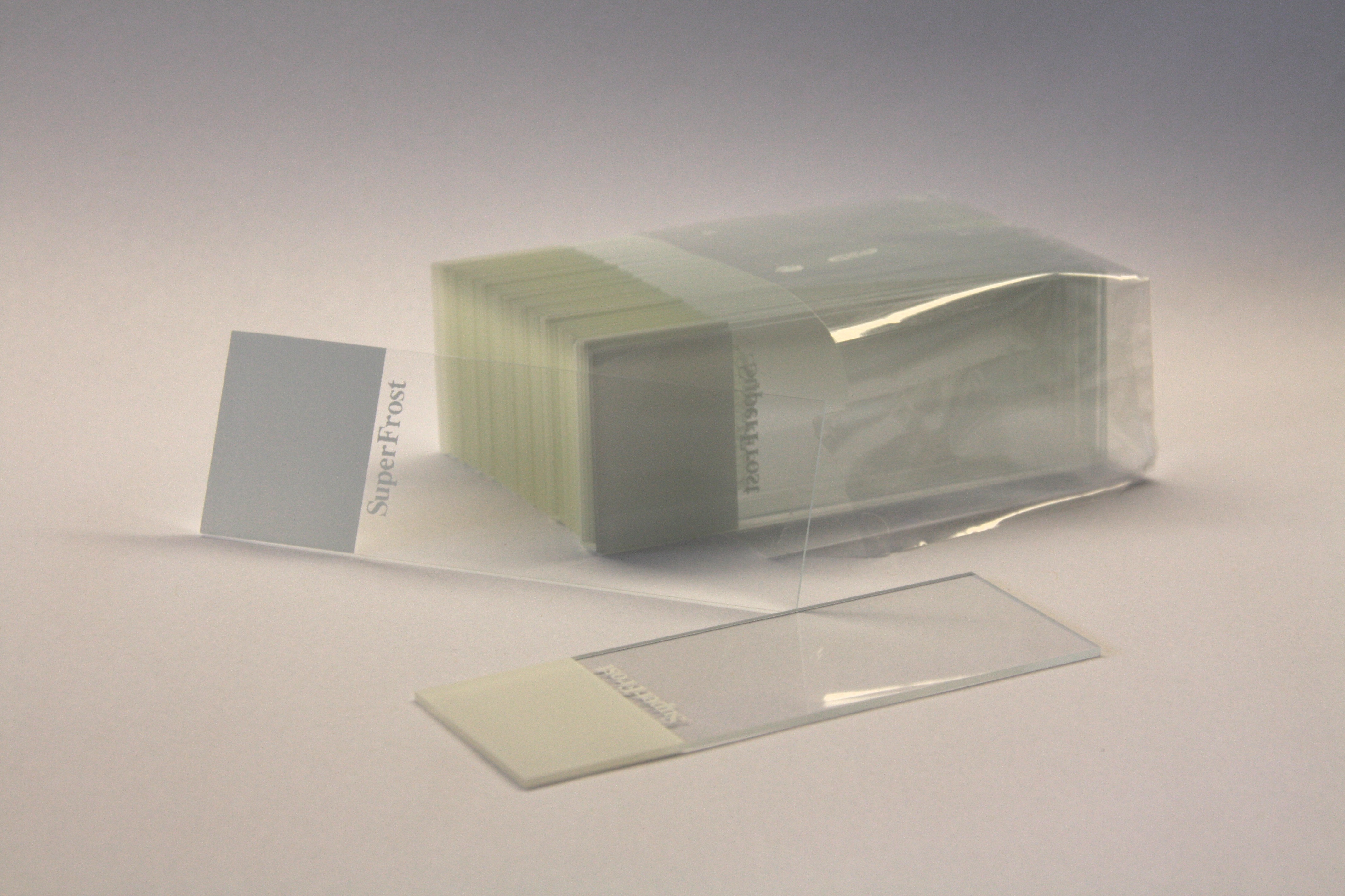
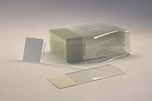 A set of standard 75 by 25 mm microscope slides. The white area can be written on to label the slide.
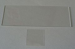 A microscope slide (top) and a cover slip (bottom)
A microscope slide is a thin flat piece of glass, typically 75 by 25 mm (3 by 1 inches) and about 1 mm thick, used to hold objects for examination under a microscope. Typically the object is placed or secured ("mounted") on the slide, and then both are inserted together in the microscope for viewing. This arrangement allows several slide-mounted objects to be quickly inserted and removed from the microscope, labeled, transported, and stored in appropriate slide cases or folders.
Microscope slides are often used together with a cover slip or cover glass, a smaller and thinner sheet of glass that is placed over the specimen. Slides are held in place on the microscope's stage by slide clips or slide clamps.
Contents
History
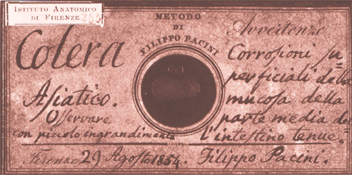
 A microscope slide prepared by Filippo Pacini in 1854, containing reference specimens from the inner mucosa of the small intestine of a cholera victim.
The origin of the concept was pieces of ivory or bone, containing specimens held between disks of transparent mica, that would slide into the gap between the stage and the objective.[citation needed] These "sliders" were popular in Victorian England until the Royal Microscopical Society introduced the standardized glass microscope slide.[citation needed]
Dimensions and types
A standard microscope slide measures about 75 mm by 25 mm (3" by 1") and is about 1 mm thick. A range of other sizes are available for various special purposes, such as 75 x 50 mm and for geological use, 46 x 27 mm for petrographic studies, and 48 x 28 mm for thin sections. Slides are usually made of common glass and their edges are often finely ground or polished.
Microscope slides are usually made of glass, such as soda lime glass or borosilicate glass, but specialty plastics are also used. Fused quartz slides are often used when ultraviolet transparency is important, e.g. in fluorescence microscopy.[1][2]
While plain slides are the most common, there are several specialized types. A concavity slide has one or more shallow depressions ("wells"), designed to hold certain samples such as liquids and tissue cultures.[3] Slides may have rounded corners for increased safety or robustness, or a cut-off corner for automated handling.[3]
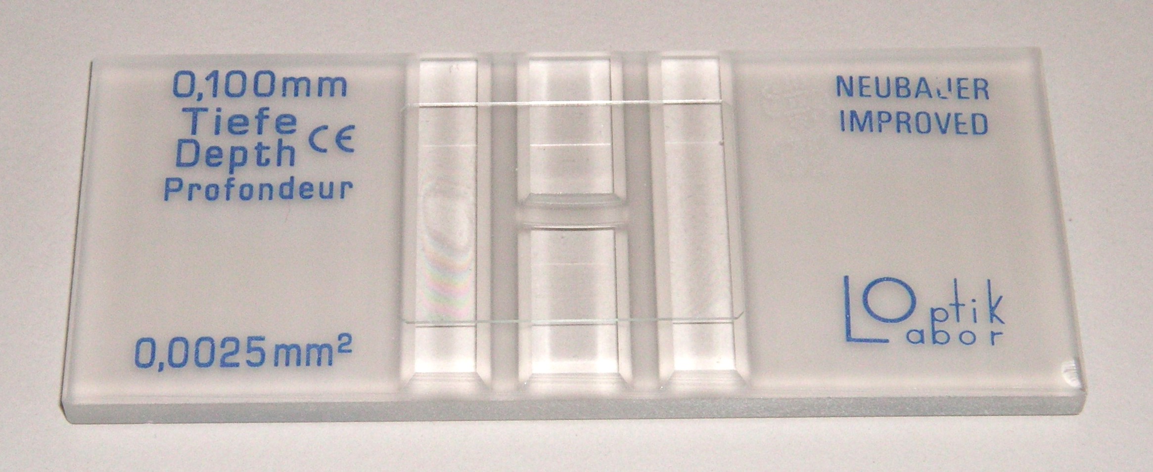
A graticule slide is marked with a grid of lines (for example, a 1 mm grid) that allows the size of objects seen under magnification to be easily estimated and provides reference areas for counting minute objects. Sometimes one square of the grid will itself be subdivided into a finer grid. Slides for specialized applications, such as cell counting, may have various reservoirs, channels and barriers etched or ground on their upper surface.[citation needed] Various permanent markings or masks may be printed, sand-blasted, or deposited on the surface by the manufacturer, usually with inert materials such as PTFE.[4]
 A Neubauer slide for cell counting.
 Microscope image of a Neubauer slide's graticule being used to count cells.
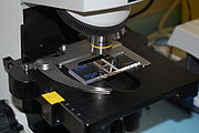 A Neubauer slide held in place on a microscope stand by a slide clamp.
Some slides have a frosted or enamel-coated area at one end, for labeling with a pencil or pen.[3] Slides may have special coatings applied by the manufacturer, e.g. for chemical inertness or enhanced cell adhesion. The coating may have a permanent electric charge to hold thin or powdery samples. Common coatings include poly-L-lysine, silanes, epoxy resins,[3][4] or even gold.[5]
Mounting
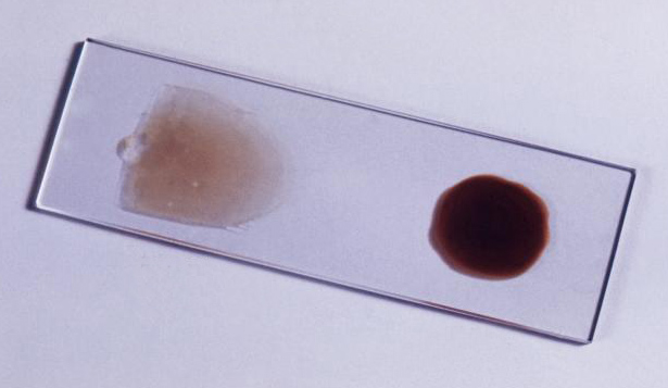
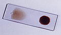 Blood smears for pathological examination, an example of wet mount.
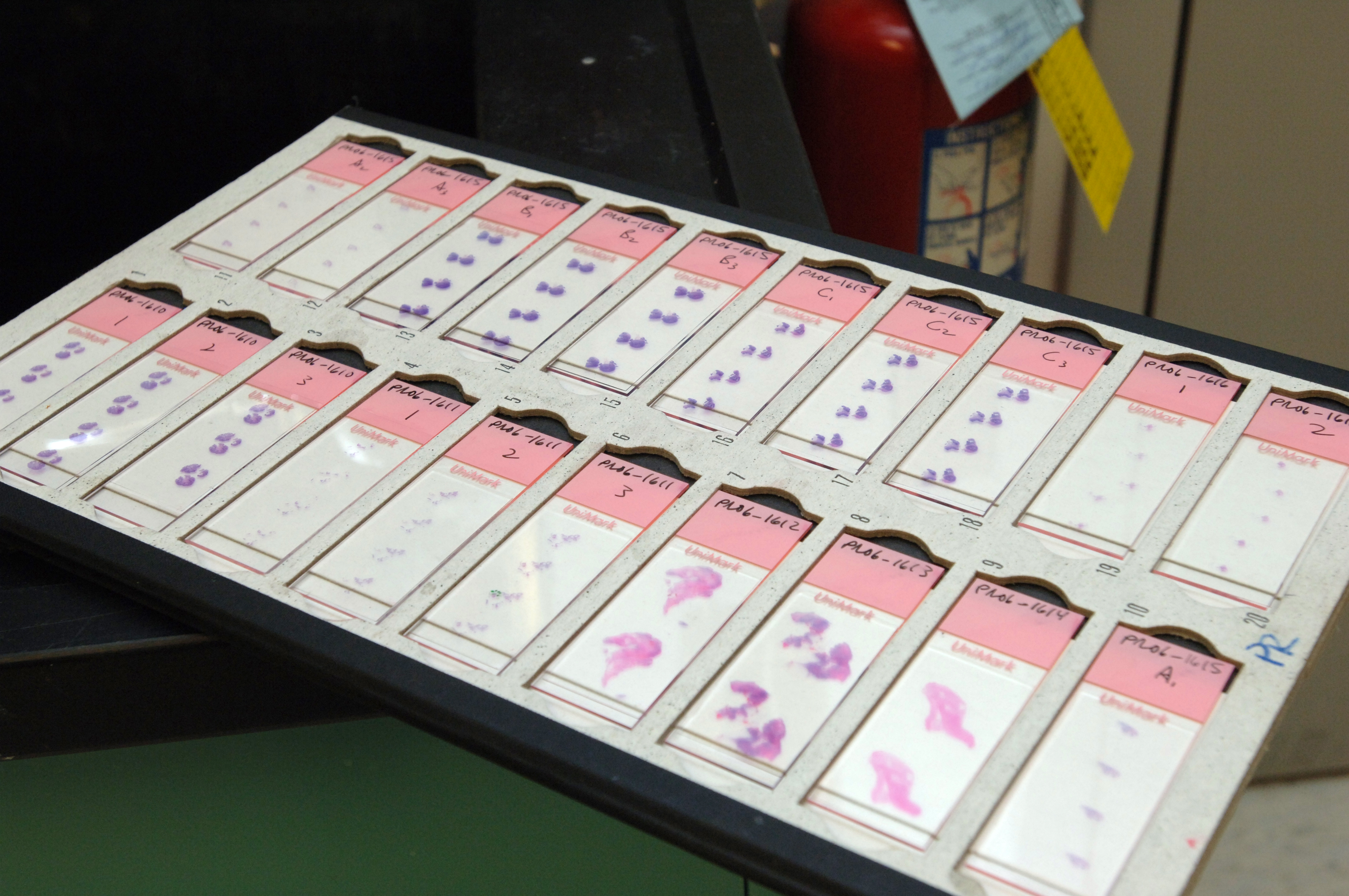
 Microscope slides with prepared, stained, and labeled tissue specimens in a standard 20-slide folder.
The mounting of specimens on microscope slides is often critical for successful viewing. The problem has been given much attention in the last two centuries and is a well-developed area with many specialized and sometimes quite sophisticated techniques.
Dry mount
In a dry mount, the simplest kind of mounting, the object is merely placed on the slide. A cover slip may be placed on top to protect the specimen and the microscope's objective and to keep the specimen still and pressed flat. This mounting can be successfully used for viewing specimens like pollen, feathers, hairs, etc. It is also used to examine particles caught in transparent membrane filters (e.g., in analysis of airborne dust).
Wet mount
In a wet mount, the specimen is placed in a drop of water or other liquid held between the slide and the cover slip by surface tension. This method is commonly used, for example, to view microscopic organisms that grow in pond water or other liquid media, especially when studying their movement and behavior. It is also used to examine physiological liquids like blood, urine, saliva, semen, and vaginal discharge. Care must be taken to exclude air bubbles that would interfere with the viewing and hamper the organisms' movements.
Prepared mount
For pathological and biological research, the specimen usually undergoes a complex histological preparation that may involve cutting it into very thin sections with a microtome, fixing it to prevent decay, removing any water contained in it, staining specific parts of it, and impregnating or infiltrating it with some transparent solid substance. As part of this process the specimen usually ends up firmly attached to the slide.
Mounting media
The mounting medium is the solution in which the specimen is embedded, generally under a cover glass. Simple liquids like water or glycerol can be considered mounting media, though the term generally refers to compounds that harden into a permanent mount. Popular mounting media include Permount, glycerol jelly, and Hoyer's mounting medium. Properties of a good mounting medium include having a refractive index close to that of glass (1.518), non-reactivity with the specimen, stability over time without crystallizing, darkening, or changing refractive index, solubility in the medium the specimen was prepared in (either aqueous or non-polar, such as xylene or toluene), and not causing the specimen stain to fade or leach.[6]
See also
References
- ^ Quartz Microscospe Slides and Cover Slips from a commercial website (Ted Pella). Accessed on 2010-01-23.
- ^ Quartz Microscope Slides and Cover Slips catalog page from a commercial website (SPI Supplies). Accessed on 2010-01-23.
- ^ a b c d Histology and Light Microscopy catalog page from a commercial website (EMS). Accessed on 2010-01-23.
- ^ a b Microscope Slides catalog page from a commercial website (TEKDON). Accessed on 2010-01-23.
- ^ Gold Coated Microscope Slides and DNA Imaging Kit catalog page from a commercial website (Asylum Research). Accessed on 2011-08-31.
- ^ Medical Laboratory Science : Theory And Practice By Ochei & Kolkatker, p. 446.
Categories:


Wikimedia Foundation. 2010.