- Cerebellar vermis
-
Brain: Cerebellar vermis 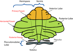
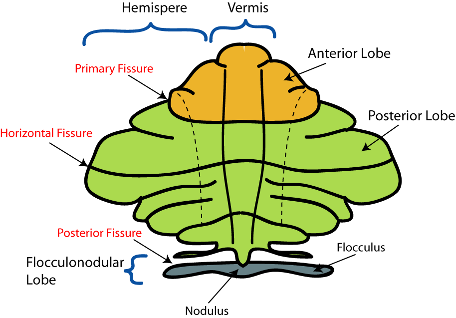
Schematic representation of the major anatomical subdivisions of the cerebellum. Superior view of an "unrolled" cerebellum, placing the vermis in one plane. 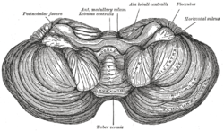
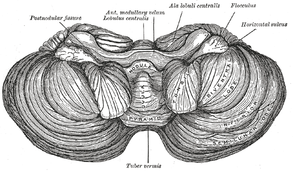
Anterior view of the cerebellum. ("Tuber vermis" labeled at bottom.) Latin vermis cerebelli Gray's subject #187 788 Part of Cerebellum NeuroNames ancil-146 NeuroLex ID birnlex_1106 Part of the structure of animal brains, the cerebellar vermis is a narrow, wormlike structure between the hemispheres of the cerebellum.
Contents
Function
It is the site of termination of the spinocerebellar pathways that carry subconscious proprioception.
Recent research on the posterior cerebellar vermis indicates that this particular area of the brain may be linked to the brain's natural ability to integrate and analyze inertial motion. Specialized cells in this area, known as Purkinje cells, are now thought to receive sensory information from the vestibular system of the inner ears and use this to compute information about the body's movement through space.[1]
There are nine subdivisons of the cerbellar vermis. They are the: lingula, central, culmen, declive, folium, tuber, pyramid, uvula, nodulus (ordered from anterior to posterior). A useful mnemonic device to remember these subdivisions is : "Like cats catching dogs for the party up north."
Clinical significance
Dandy Walker malformation is a congenital brain malformation that is characterized by enlarged posterior fossa and in which the cerebellar vermis is absent or present in merely a rudimentary form. It is also commonly associated with dysplasias of brainstem nuclei.
See also
- cerebellar vermis hypoplasia, a genetic ciliopathy
Additional images
-

Human brain midsagittal view
References
External links
- Photo - rollover to see highlighted at University of Texas Southwestern Medical Center at Dallas
- Diagram at medfriendly.com
- BrainMaps at UCDavis Vermis
Human brain, rhombencephalon, metencephalon: cerebellum (TA 14.1.07, GA 9.788) Surface anatomy LobesMedial/lateralVermis: anterior (Central lobule, Culmen, Lingula) · posterior (Folium, Tuber, Uvula) · Vallecula of cerebellum
Hemisphere: anterior (Alar central lobule) · posterior (Biventer lobule, Cerebellar tonsil)Grey matter Molecular layer (Stellate cell, Basket cell)
Purkinje cell layer (Purkinje cell, Bergmann glia cell = Golgi epithelial cell)
Granule cell layer (Golgi cell, Granule cell, Unipolar brush cell)
Fibers: Mossy fibers · Climbing fiber · Parallel fiberWhite matter InternalPedunclesInferior (medulla): Dorsal spinocerebellar tract · Olivocerebellar tract · Cuneocerebellar tract · Juxtarestiform body (Vestibulocerebellar tract)
Middle (pons): Pontocerebellar fibers
Superior (midbrain): Ventral spinocerebellar tract · Dentatothalamic tract · Trigeminocerebellar fibersCategories:- Cerebellum
- Neuroanatomy stubs

Wikimedia Foundation. 2010.