- Complement receptor 2
-
Complement component (3d/Epstein Barr virus) receptor 2, also known as CR2 and CD21, is a protein involved in the complement system. It binds to iC3b (inactive derivative of C3b), C3dg, or C3d.[1] B cells have CR2 receptors on their surfaces, allowing the complement system to play a role in B-cell activation and maturation [2]
Contents
Interactions
CR2 on mature B cells form a complex with two other membrane proteins, CD19 and CD81(=TAPA-1). The CR2-CD19-CD81 complex is often called the B cell coreceptor complex,[3] because CR2 binds to antigens through attached C3d (or iC3b or C3dg) when the membrane IgM binds to the antigen. This results in the B cell having greatly enhanced response to the antigen.[1]
Complement receptor 2 has been shown to interact with CD19.[4][5]
Epstein-Barr virus (EBV) binds to B cells at CR2 during infection of these cells. Yefenof et al. (1976) found complete overlapping of EBV receptors and C3 receptors on human B cells.[2][6]
Immunohistochemistry
Although CR2 is present on all mature B-cells and follicular dendritic cells (FDCs), this only readily apparent when immunohistochemistry is performed on frozen sections. In more conventional paraffin-embedded tissue samples, only the follicular dendritic cells retain the staining pattern. As a result, CR2, more commonly called CD21 in the context of immunohistochemistry, can be used to demonstrate the FDC meshwork in lymphoid tissue.
This feature can be useful in examining tissue where the normal germinal centres have been effaced by disease processes, such as HIV infection. The pattern of the FDC meshwork may also be altered in some neoplastic conditions, such as B-cell MALT lymphomas, mantle cell lymphoma, and some T cell lymphomas. Castleman's disease is typified by the presence of abnormal FDCs, and both this, and malignant FDC tumours may therefore be demonstrated using CR2/CD21 antibodies.[7]
References
- ^ a b Frank K, Atkinson JP (2001). "Complement system." In Austen KF, Frank K, Atkinson JP, Cantor H. eds. Samter's Immunologic Diseases, 6th ed. Vol. 1, p. 281-298, Philadelphia: Lippincott Williams & Wilkins, ISBN 0781721202.
- ^ a b "Entrez Gene: CR2 complement component (3d/Epstein Barr virus) receptor 2". http://www.ncbi.nlm.nih.gov/sites/entrez?Db=gene&Cmd=ShowDetailView&TermToSearch=1380.
- ^ Abbas AK, Lichtman AH (2003). Cellular and Molecular Immunology, 5th ed. Philadelphia: Saunders, ISBN 0-7216-0008-5
- ^ Bradbury, L E; Kansas G S, Levy S, Evans R L, Tedder T F (Nov. 1992). "The CD19/CD21 signal transducing complex of human B lymphocytes includes the target of antiproliferative antibody-1 and Leu-13 molecules". J. Immunol. (UNITED STATES) 149 (9): 2841–50. ISSN 0022-1767. PMID 1383329.
- ^ Horváth, G; Serru V, Clay D, Billard M, Boucheix C, Rubinstein E (Nov. 1998). "CD19 is linked to the integrin-associated tetraspans CD9, CD81, and CD82". J. Biol. Chem. (UNITED STATES) 273 (46): 30537–43. doi:10.1074/jbc.273.46.30537. ISSN 0021-9258. PMID 9804823.
- ^ Yefenof E, Klein G, Jondal M, Oldstone MB (June 1976). "Surface markers on human B and T-lymphocytes. IX. Two-color immunofluorescence studies on the association between ebv receptors and complement receptors on the surface of lymphoid cell lines". Int. J. Cancer 17 (6): 693–700. doi:10.1002/ijc.2910170602. PMID 181330.
- ^ Leong, Anthony S-Y; Cooper, Kumarason; Leong, F Joel W-M (2003). Manual of Diagnostic Cytology (2 ed.). Greenwich Medical Media, Ltd.. pp. 93–94. ISBN 1-84110-100-1.
Further reading
- Cooper NR, Bradt BM, Rhim JS, Nemerow GR (1990). "CR2 complement receptor.". J. Invest. Dermatol. 94 (6 Suppl): 112S?117S. doi:10.1111/1523-1747.ep12876069. PMID 2161885.
- Gauffre A, Viron A, Barel M, et al. (1992). "Nuclear localization of the Epstein-Barr virus/C3d receptor (CR2) in the human Burkitt B lymphoma cell, Raji.". Mol. Immunol. 29 (9): 1113–20. doi:10.1016/0161-5890(92)90044-X. PMID 1323059.
- Levy E, Ambrus J, Kahl L, et al. (1992). "T lymphocyte expression of complement receptor 2 (CR2/CD21): a role in adhesive cell-cell interactions and dysregulation in a patient with systemic lupus erythematosus (SLE).". Clin. Exp. Immunol. 90 (2): 235–44. doi:10.1111/j.1365-2249.1992.tb07935.x. PMC 1554594. PMID 1424280. http://www.pubmedcentral.nih.gov/articlerender.fcgi?tool=pmcentrez&artid=1554594.
- Matsumoto AK, Kopicky-Burd J, Carter RH, et al. (1991). "Intersection of the complement and immune systems: a signal transduction complex of the B lymphocyte-containing complement receptor type 2 and CD19.". J. Exp. Med. 173 (1): 55–64. doi:10.1084/jem.173.1.55. PMC 2118751. PMID 1702139. http://www.pubmedcentral.nih.gov/articlerender.fcgi?tool=pmcentrez&artid=2118751.
- Tuveson DA, Ahearn JM, Matsumoto AK, Fearon DT (1991). "Molecular interactions of complement receptors on B lymphocytes: a CR1/CR2 complex distinct from the CR2/CD19 complex.". J. Exp. Med. 173 (5): 1083–9. doi:10.1084/jem.173.5.1083. PMC 2118840. PMID 1708808. http://www.pubmedcentral.nih.gov/articlerender.fcgi?tool=pmcentrez&artid=2118840.
- Kalli KR, Ahearn JM, Fearon DT (1991). "Interaction of iC3b with recombinant isotypic and chimeric forms of CR2.". J. Immunol. 147 (2): 590–4. PMID 1830068.
- Barel M, Gauffre A, Lyamani F, et al. (1991). "Intracellular interaction of EBV/C3d receptor (CR2) with p68, a calcium-binding protein present in normal but not in transformed B lymphocytes.". J. Immunol. 147 (4): 1286–91. PMID 1831222.
- Delcayre AX, Salas F, Mathur S, et al. (1991). "Epstein Barr virus/complement C3d receptor is an interferon alpha receptor.". EMBO J. 10 (4): 919–26. PMC 452735. PMID 1849076. http://www.pubmedcentral.nih.gov/articlerender.fcgi?tool=pmcentrez&artid=452735.
- Kurtz CB, Paul MS, Aegerter M, et al. (1989). "Murine complement receptor gene family. II. Identification and characterization of the murine homolog (Cr2) to human CR2 and its molecular linkage to Crry.". J. Immunol. 143 (6): 2058–67. PMID 2528587.
- Fujisaku A, Harley JB, Frank MB, et al. (1989). "Genomic organization and polymorphisms of the human C3d/Epstein-Barr virus receptor.". J. Biol. Chem. 264 (4): 2118–25. PMID 2563370.
- Moore MD, Cooper NR, Tack BF, Nemerow GR (1988). "Molecular cloning of the cDNA encoding the Epstein-Barr virus/C3d receptor (complement receptor type 2) of human B lymphocytes.". Proc. Natl. Acad. Sci. U.S.A. 84 (24): 9194–8. doi:10.1073/pnas.84.24.9194. PMC 299719. PMID 2827171. http://www.pubmedcentral.nih.gov/articlerender.fcgi?tool=pmcentrez&artid=299719.
- Weis JJ, Toothaker LE, Smith JA, et al. (1988). "Structure of the human B lymphocyte receptor for C3d and the Epstein-Barr virus and relatedness to other members of the family of C3/C4 binding proteins.". J. Exp. Med. 167 (3): 1047–66. doi:10.1084/jem.167.3.1047. PMC 2188894. PMID 2832506. http://www.pubmedcentral.nih.gov/articlerender.fcgi?tool=pmcentrez&artid=2188894.
- Weis JJ, Fearon DT, Klickstein LB, et al. (1986). "Identification of a partial cDNA clone for the C3d/Epstein-Barr virus receptor of human B lymphocytes: homology with the receptor for fragments C3b and C4b of the third and fourth components of complement.". Proc. Natl. Acad. Sci. U.S.A. 83 (15): 5639–43. doi:10.1073/pnas.83.15.5639. PMC 386344. PMID 3016712. http://www.pubmedcentral.nih.gov/articlerender.fcgi?tool=pmcentrez&artid=386344.
- Weis JH, Morton CC, Bruns GA, et al. (1987). "A complement receptor locus: genes encoding C3b/C4b receptor and C3d/Epstein-Barr virus receptor map to 1q32.". J. Immunol. 138 (1): 312–5. PMID 3782802.
- Aubry JP, Pochon S, Gauchat JF, et al. (1994). "CD23 interacts with a new functional extracytoplasmic domain involving N-linked oligosaccharides on CD21.". J. Immunol. 152 (12): 5806–13. PMID 7515913.
- Matsumoto AK, Martin DR, Carter RH, et al. (1993). "Functional dissection of the CD21/CD19/TAPA-1/Leu-13 complex of B lymphocytes.". J. Exp. Med. 178 (4): 1407–17. doi:10.1084/jem.178.4.1407. PMC 2191213. PMID 7690834. http://www.pubmedcentral.nih.gov/articlerender.fcgi?tool=pmcentrez&artid=2191213.
- Barel M, Balbo M, Gauffre A, Frade R (1995). "Binding sites of the Epstein-Barr virus and C3d receptor (CR2, CD21) for its three intracellular ligands, the p53 anti-oncoprotein, the p68 calcium binding protein and the nuclear p120 ribonucleoprotein.". Mol. Immunol. 32 (6): 389–97. doi:10.1016/0161-5890(95)00005-Y. PMID 7753047.
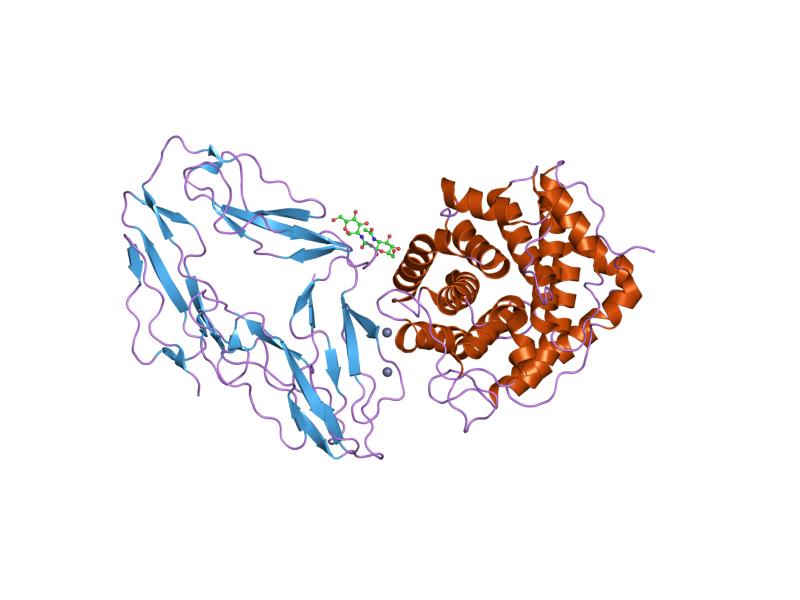
PDB gallery 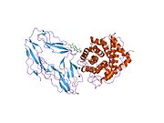 1ghq: CR2-C3D COMPLEX STRUCTURE
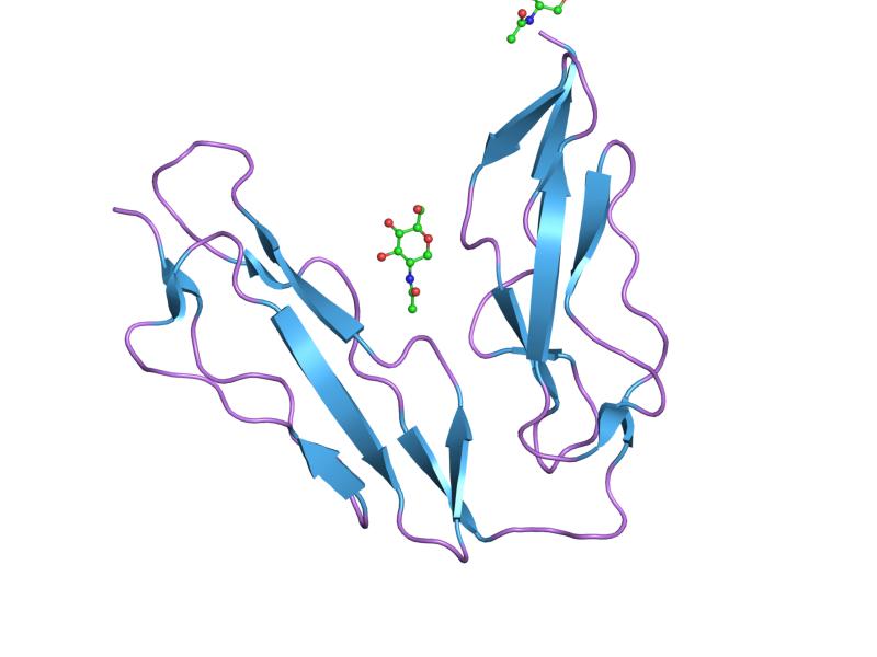
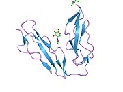 1ly2: Crystal structure of unliganded human CD21 SCR1-SCR2 (Complement receptor type 2)
External links
Proteins: complement system (C, L, A) Activators/enzymes EarlyMiddleLateInhibitors Complement receptors 1-50 CD1 (a-c, 1A, 1D, 1E) · CD2 · CD3 (γ, δ, ε) · CD4 · CD5 · CD6 · CD7 · CD8 (a) · CD9 · CD10 · CD11 (a, b, c) · CD13 · CD14 · CD15 · CD16 (A, B) · CD18 · CD19 · CD20 · CD21 · CD22 · CD23 · CD24 · CD25 · CD26 · CD27 · CD28 · CD29 · CD30 · CD31 · CD32 (A, B) · CD33 · CD34 · CD35 · CD36 · CD37 · CD38 · CD39 · CD40 · CD41 · CD42 (a, b, c, d) · CD43 · CD44 · CD45 · CD46 · CD47 · CD48 · CD49 (a, b, c, d, e, f) · CD5051-100 CD51 · CD52 · CD53 · CD54 · CD55 · CD56 · CD57 · CD58 · CD59 · CD61 · CD62 (E, L, P) · CD63 · CD64 (A, B, C) · CD66 (a, b, c, d, e, f) · CD68 · CD69 · CD70 · CD71 · CD72 · CD73 · CD74 · CD78 · CD79 (a, b) · CD80 · CD81 · CD82 · CD83 · CD84 · CD85 (a, d, e, h, j, k) · CD86 · CD87 · CD88 · CD89 · CD90 · CD91- CD92 · CD93 · CD94 · CD95 · CD96 · CD97 · CD98 · CD99 · CD100101-150 CD101 · CD102 · CD103 · CD104 · CD105 · CD106 · CD107 (a, b) · CD108 · CD109 · CD110 · CD111 · CD112 · CD113 · CD114 · CD115 · CD116 · CD117 · CD118 · CD119 · CD120 (a, b) · CD121 (a, b) · CD122 · CD123 · CD124 · CD125 · CD126 · CD127 · CD129 · CD130 · CD131 · CD132 · CD133 · CD134 · CD135 · CD136 · CD137 · CD138 · CD140b · CD141 · CD142 · CD143 · CD144 · CD146 · CD147 · CD148 · CD150151-200 CD151 · CD152 · CD153 · CD154 · CD155 · CD156 (a, b, c) · CD157 · CD158 (a, d, e, i, k) · CD159 (a, c) · CD160 · CD161 · CD162 · CD163 · CD164 · CD166 · CD167 (a, b) · CD168 · CD169 · CD170 · CD171 · CD172 (a, b, g) · CD174 · CD177 · CD178 · CD179 (a, b) · CD181 · CD182 · CD183 · CD184 · CD185 · CD186 · CD191 · CD192 · CD193 · CD194 · CD195 · CD196 · CD197 · CDw198 · CDw199 · CD200201-250 CD201 · CD202b · CD204 · CD205 · CD206 · CD207 · CD208 · CD209 · CDw210 (a, b) · CD212 · CD213a (1, 2) · CD217 · CD218 (a, b) · CD220 · CD221 · CD222 · CD223 · CD224 · CD225 · CD226 · CD227 · CD228 · CD229 · CD230 · CD233 · CD234 · CD235 (a, b) · CD236 · CD238 · CD239 · CD240CE · CD240D · CD241 · CD243 · CD244 · CD246 · CD247- CD248 · CD249251-300 CD252 · CD253 · CD254 · CD256 · CD257 · CD258 · CD261 · CD262 · CD264 · CD265 · CD266 · CD267 · CD268 · CD269 · CD271 · CD272 · CD273 · CD274 · CD275 · CD276 · CD278 · CD279 · CD280 · CD281 · CD282 · CD283 · CD284 · CD286 · CD288 · CD289 · CD290 · CD292 · CDw293 · CD294 · CD295 · CD297 · CD298 · CD299301-350 Lymphoid Pre-B cell: CD10/CALLA · CD79A
mature: CD19 · CD20 · CD21/CR2 · CD23/FcεRII · CD127 · CD40
plasma cell: CD38 · CD138T/NKNK cellAllAllMyeloid CFU-GM/
MyelomonocyteCFU-MegCFU-ECD36 · CD71All (pan-myeloid)Stem cell Transmembrane receptors: Immunoglobulin superfamily immune receptors Antibody receptor:
Fc receptorSecretoryAntigen receptor Antigen receptorAccessory moleculesT cellsAntigen receptorAccessory moleculesCytokine receptor see cytokine receptorsKiller-cell IG-like receptors Leukocyte IG-like receptors B trdu: iter (nrpl/grfl/cytl/horl), csrc (lgic, enzr, gprc, igsr, intg, nrpr/grfr/cytr), itra (adap, gbpr, mapk), calc, lipd; path (hedp, wntp, tgfp+mapp, notp, jakp, fsap, hipp, tlrp)Categories:- Human proteins
- Complement system
- Chromosome 1 gene stubs


Wikimedia Foundation. 2010.